Image Encryption and Watermarking Using DWT Matlab Project With Source Code | Final Year Project Code

ABSTRACT

          The use of Internet technology has led to the availability of different multimedia data in various formats. The unapproved customers misuse multimedia information by conveying them on various web objections to acquire cash deceptively without the first copyright holder’s intervention. Due to the rise in cases of COVID-19, lots of patient information are leaked without their knowledge, so an intelligent technique is required to protect the integrity of patient data by placing an invisible signal known as a watermark on the medical images. In this project encryption and watermarking is proposed using discrete wavelet transform algorithm on both standard and medical images. The project addresses the use of digital rights management in medical field applications such as encrypting and embedding the watermark in medical images. The various quality parameters are used to figure out the evaluation of the developed method. This project is developed in matlab.
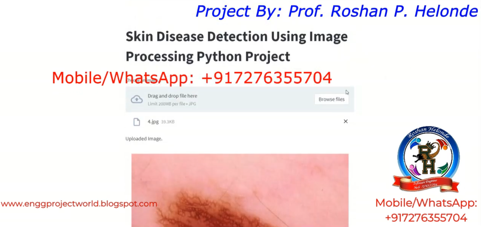
PROJECT OUTPUT


PROJECT DEMO VIDEO

Contact:
Prof. Roshan P. Helonde
Mobile: +91-7276355704
WhatsApp: +91-7276355704
Email: roshanphelonde@rediffmail.com
Share:

Bone Fracture Detection Using Image Processing Python Project With Source Code | Final Year Project Code

ABSTRACT

         Analysis of medical images plays a very important role in clinical decision making. For a long time it has required extensive involvement of a human expert. However, recent progress in data mining techniques, especially in machine learning, allows for creating decision models and support systems that help to automatize this task and provide clinicians with patient-specific therapeutic and diagnostic suggestions. In this project, we describe a study aimed at building a decision model (a classifier) that would predict the type of treatment (surgical vs. non-surgical) for patients with bone fractures based on their X-ray images. We consider two types of features extracted from images (structural and textural) and used them to construct multiple classifiers that are later evaluated in a computational experiment. Structural features are computed by applying the Hough transform, while textural information is obtained from Gray-level occurrence matrix. In research reported by other authors structural and textural features were typically considered separately. Our findings show that while structural features have better predictive capabilities, they can benefit from combining them with textural ones. This project is developed in python.

PROJECT OUTPUT


PROJECT DEMO VIDEO

Contact:
Prof. Roshan P. Helonde
Mobile: +91-7276355704
WhatsApp: +91-7276355704
Email: roshanphelonde@rediffmail.com
Share:

Medical Image Encryption Decryption Matlab Project With Source Code | Medical Data Encryption and Decryption

 ABSTRACT

             In recent years mostly all the health centers and hospitals use the wireless networks and internet for biomedical information exchanging, the secure of this information in not verified and cannot be grantee in such environment, the personality of patient and for security concerns inside such institutions there is a need for encryption system that can easily encrypt the biomedical data and it can beshared with other centers via internet without and concerns about privacy. Our system based on advanced encryption standard with encryption and decryption taking to consideration the criticality of data that been encrypted. Medical image security is very important issue in new world technologies with the internet of things revolution everything is connected to the internet and need to protected and authenticated, our project can encrypt the medical images for popular people or the critical situation patient that can help to protect the patients privacy by merging many techniques.

PROJECT OUTPUT


PROJECT DEMO VIDEO

Contact:
Prof. Roshan P. Helonde
Mobile: +91-7276355704
WhatsApp: +917276355704
Email: roshanphelonde@rediffmail.com
Share:

Skin Disease Detection Using Image Processing CNN | Skin Disease Classification Using CNN Python Project With Source Code

ABSTRACT

              Skin diseases are hazardous and often contagious, especially melanoma, eczema, and impetigo. These skin diseases can be cured if detected early. The fundamental problem with it is, only an expert dermatologist is able to detect and classify such disease. Sometimes, the doctors also fail to correctly classify the disease and hence provide inappropriate medications to the patient. Our research proposes a skin disease detection method based on CNN Image Processing Techniques. Our system is Personal Computer based so can be used even in remote areas. The patient needs to provide the image of the infected area and it is given as an input to the application. Image Processing cnn techniques process it and deliver the accurate output.

PROJECT OUTPUT


PROJECT DEMO VIDEO

Contact:
Prof. Roshan P. Helonde
Mobile: +917276355704
WhatsApp: +917276355704
Email: roshanphelonde@rediffmail.com
Share:

Fingernail Disease Detection Using Image Processing Matlab Project Source Code | Final Year Project Code

ABSTRACT

          This project gives idea to predict diseases using the color of the nail at early stage of diagnosis. The main aim of our project is to analyze the disease without causing harm to humans. In earlier traditional system of disease detection, doctors observe the nails of patients and will predict the disease. Many diseases can be identified by analyzing nails of patients. But it is difficult for human eyes to differentiate the slight changes in color. So it is less accurate and time consuming. Our proposed system can be quite useful to overcome this issue since it is fully computer based. The input to the proposed system is image of nail. The system will process the nail image and will extract the nail’s features to diagnose the disease. Human nail consist of various features, our proposed system uses nail color changes to diagnose the disease. Here, first training set data is prepared from nail images of patients with specific diseases. This training data set is compared with extracted feature from input nail image to obtain the result. In our experiment, we found that training set data are correctly matched with color feature of nail image results. It is focused on the system of image recognition on the basis of color analysis. The proposed system is based on the algorithm which automatically extracts only nails area from scanned back side of palm (Region of Interest). These selected pixels are processed for further analysis using median filters. The system is fully computer based, so even small discontinuities in color values are observed, and we can detect color changes in the initial stage of disease.

PROJECT OUTPUT


PROJECT DEMO VIDEO

Contact:
Prof. Roshan P. Helonde
Mobile: +91-7276355704
WhatsApp: +917276355704
Email: roshanphelonde@rediffmail.com
Share:

Alzheimer's Detection Using Image Processing | Alzheimers Disease Detection Matlab Project With Source Code | Final Year Project

ABSTRACT

            Early detection of Alzheimer's disease (AD) is important so that preventative measures can be taken. Current techniques for detecting AD rely on cognitive impairment testing which unfortunately does not yield accurate diagnoses until the patient has progressed beyond a moderate AD. Alzheimer's disease considered being one of the acute diseases that cause the human death especially in people above 60 years old. Many computer-aided diagnosis systems are now widely spread to aid in Alzheimer diagnosis. Therefore, an automated and reliable computer-aided diagnostic system for diagnosing and classifying the brain diseases has been proposed. MRI (Magnetic resonance Imaging) is one source of brain diseases detection tools, but using MRI in Alzheimer classification is considered to be difficult process according to the variance and complexity of brain tissue. This project presents a survey of the most famous techniques used for the classification of brain diseases based on MRI. The Alzheimer detection and classification systems consist of four stages, namely, MRI preprocessing, Segmentation, Feature extraction, and Classification respectively. In the first stage, the main task is to eliminate the medical resonance images (MRI) noise which may cause due to light reflections or any inaccuracies in the imaging medium. The second stage, which is the stage where the region of interest is extracted (Alzheimer region). In the third stage, the features related to MRI images will be obtained and stored in an image vector to be ready for the classification process and finally the fourth stages, where classifier will take place to specify the Alzheimer kind.

PROJECT OUTPUT


PROJECT DEMO VIDEO

Contact:
Prof. Roshan P. Helonde
Mobile: +91-7276355704
WhatsApp: +917276355704
Email: roshanphelonde@rediffmail.com
Share:

Lossless Image Compression Using Huffman Coding | Huffman Image Compression Matlab Project Source Code

 ABSTRACT

            The lossless compression is that allows the original data to be perfectly reconstructed from the compressed data. Lossless compression programs do two things in sequence: the first step generates a statistical model for the input data, and the second step uses this model to map input data to bit sequences in such a way that probable. The main objective of image compression is to decrease the redundancy of the image data which helps in increasing the capacity of storage and efficient transmission. Image compression aids in decreasing the size in bytes of a digital image without degrading the quality of the image to an undesirable level. Image compression plays an important role in computer storage and transmission. The purpose of data compression is that we can reduce the size of data to save storage and reduce time for transmission. Image compression is a result of applying data compression to the digital image.

PROJECT OUTPUT


PROJECT DEMO VIDEO

Contact:
Prof. Roshan P. Helonde
Mobile: +91-7276355704
WhatsApp: +917276355704
Email: roshanphelonde@rediffmail.com
Share:

Fruit Disease Detection Using Image Processing Python Project With Source Code | Fruit Disease Identification Using Python Project

 ABSTRACT

            Diseases in fruit cause devastating problem in economic losses and production in agricultural industry worldwide. In this project, a solution for the detection and classification of fruit diseases is proposed and experimentally validated. Our experimental results express that the proposed solution can significantly support accurate detection and automatic classification of fruit diseases. An early detection of fruit diseases can aid in decreasing such losses and can stop further spread of diseases. A lot of work has been done to automate the visual inspection of the fruits by machine vision with respect to size and color. However, detection of defects in the fruits using images is still problematic due to the natural variability of skin color in different types of fruits, high variance of defect types, and presence of stem/calyx. To know what control factors to consider next year to overcome similar losses, it is of great significance to analyze what is being observed. This project is developed in python.

PROJECT OUTPUT


PROJECT DEMO VIDEO

Contact:
Prof. Roshan P. Helonde
Mobile: +91-7276355704
WhatsApp: +917276355704
Email: roshanphelonde@rediffmail.com
Share:

Skin Disease Prediction Using CNN Python Project With Source Code | Skin Disease Classfication Using CNN

 ABSTRACT

         Skin cancer also known as melanoma it is one of the deadliest form of cancer if not recognized in time. Since the pigmented areas/moles of the skin can be nicely observed by simple, non-invasive visual inspection the clinical protocols of its recognition also consider several visual features. Melanoma is the deadliest form of skin cancer, which is considered one of the most common human malignancies in the world. Early detection of this disease can affect the result of the illness and improve the chance of surviving. The tremendous improvement of deep learning algorithms in image recognition tasks promises a great success for medical image analysis, in particular, melanoma classification for skin cancer diagnosis. Activation functions play an important role in the performance of deep neural networks for image recognition problems as well as medical image classification. Melanin is the pigment that discerns the color of human skin. The special cells produce melanin in the skin. If these cells are damaged or unhealthy, skin discoloration is visible. Skin pigment discoloration on cheeks is a hazardous fact as a symptom of human skin disease with a possibility of losing natural beauty. The extracted information of the skin discoloration can work as a guide to diagnosis the disease. In this research, different imaging techniques like preprocessing method, segmentation and morphological operations are used to analyze and extract the information of cheek’s discoloration lesion by measuring the area of lesion on skin.

PROJECT OUTPUT


PROJECT DEMO VIDEO

Contact:
Prof. Roshan P. Helonde
Mobile: +91-7276355704
WhatsApp: +91-7276355704
Email: roshanphelonde@rediffmail.com
Share:

Total Pageviews

CONTACT US

Prof. Roshan P. Helonde
Mobile: +917276355704
WhatsApp: +917276355704
Email: roshanphelonde@rediffmail.com

Enter Project Title

Popular Projects

All Archive

Contact Form

Name

Email *

Message *