Showing posts with label Biomedical Projects. Show all posts

Melanoma Skin Cancer Detection Using Python Opencv | Skin Cancer Prediction Using CNN | Final Year Major Project

ABSTRACT

                Many of the skin diseases are very dangerous, particularly if not treated at an early stage. Skin diseases are becoming common because of the increasing pollution. Skin diseases tend to pass from one person to another. Human habits tend to assume that some Melanoma Skin Cancer are not serious problems. Sometimes, most of the people try to treat these infections of the skin using their own method. However, if these treatments are not suitable for that particular skin problem then it would make it worse. And also sometimes they may not be aware of the dangerous of their Melanoma Skin Cancer, for instance skin cancers. With advance of medical imaging technologies, the acquired data information is getting so rich toward beyond the human’s capability of visual recognition and efficient use for clinical assessment. In this project we propose a diagnosis system which will enable users to detect and recognize skin diseases with the help of image processing and provide the user advises or treatments based on the results obtained in a shorter time period than the existing methods. In this project, we will be constructing a diagnosis system based on the techniques of Image Processing. We will be making use of Python to perform the pre-processing and processing of the skin images of the users. This processing will be conducted on the different skin patterns and will be analyzed to obtain the results from which we can identify which skin disease the user is suffering from. This data will help in early detection of the skin diseases and in providing their cure. Through this we will be finding a cost effective and feasible test method for the detection of skin disorders. The results obtained will be classified according to the given prototype and diagnosis accuracy assessment will be performed to provide users with efficient and fast results. This project is developed in python.

PROJECT OUTPUT


PROJECT DEMO VIDEO

Contact:
Prof. Roshan P. Helonde
Mobile: +91-7276355704
WhatsApp: +91-7276355704
Email: roshanphelonde@rediffmail.com
Share:

Palmprint Recognition Using Image Processing | Palmprint Detection Using Matlab Project With Source Code | Final Year Major Project

ABSTRACT

                 Palm  print  authentication  is  one  of  the  modern  bio-metric techniques, which employs the vein pattern  in  the  human palm  to  verify  the  person.  The merits  of  palm  vein  on classical  bio-metric  (e.g.  fingerprint,  iris,  face)  are  a  low risk  of  falsification,  difficulty  of  duplicated  and  stability. In  this  Project,  a  new  method  is  proposed  for  personal verification  based  on  palm  Print  features.  In  the propose method,  the  palm  vein  images  are  firstly  enhanced  and then  the  features  are extracted  by  using  bank  of  Gabor filters. Bio-metric   technology   refers   to   a pattern recognition system  which  depends  on  physical  or  behavioral  features for the  person  identification. This project is developed in matlab using image processing. Palmprint Recognition Using Image Processing. Palmprint Detection Using Matlab Project With Source Code.

PROJECT OUTPUT


PROJECT DEMO VIDEO

Contact:
Prof. Roshan P. Helonde
Mobile: +91-7276355704
WhatsApp: +91-7276355704
Email: roshanphelonde@rediffmail.com
Share:

Brain Tumor Detection Using Segmentation and KNN Clustering | Brain Tumor Detection Using Matlab | Major Project with Source Code

 ABSTRACT

            Image processing is a process where input image is processed to get output also as an image or attributes of the image. Main aim of all image processing techniques is to recognize the image or object under consideration easier visually. Segmentation of images holds a crucial position in the field of image processing. In medical imaging, segmentation is important for feature extraction, image measurements and image display. A tumor can be defined as a mass which grows without any control of normal forces. Real time diagnosis of tumors by using more reliable algorithms has been an active of the latest developments in medical imaging and detection of brain tumor in MR and CT scan images. Hence image segmentation is the fundamental problem used in tumor detection. Image segmentation can be defined as the partition or segmentation of a digital image into similar regions with a main aim to simplify the image under consideration into something that is more meaningful and easier to analyze visually.

         Brain tumor is an abnormal growth caused by cells reproducing themselves in an uncontrolled manner. Magnetic Resonance Imager (MRI) is the commonly used device for diagnosis. In MR images, the amount of data is too much for manual interpretation and analysis. During the past few years, brain tumor segmentation in Magnetic Resonance Imaging(MRI) has become an emergent research area in the field of medical imaging system. Accurate detection of size and location of brain tumor plays a vital role in the diagnosis of tumor. Image processing is an active research area in which medical image processing is a highly challenging field. Image segmentation plays a significant role in image processing as it helps in the extraction of suspicious regions from the medical images. In this project an efficient algorithm is proposed for tumor detection based on segmentation of brain MRI images using KNN clustering. This project is developed in matlab.

PROJECT OUTPUT


PROJECT DEMO VIDEO

Contact:
Prof. Roshan P. Helonde
Mobile: +917276355704
WhatsApp: +917276355704
Email: roshanphelonde@rediffmail.com
Share:

Skin Cancer Detection Using Deep Learning CNN Matlab Project Code Final Year IEEE Project

  ABSTRACT

          Skin cancer is a widespread, global, and potentially deadly disease, which over the last three decades has afflicted more lives in the USA than all other forms of cancer combined. There have been a lot of promising recent works utilizing deep network architectures for developing automated skin lesion segmentation. Melanin is the pigment that discerns the color of human skin. The special cells produce melanin in the skin. If these cells are damaged or unhealthy, skin discoloration is visible. Skin pigment discoloration is a hazardous fact as a symptom of human skin cancer with a possibility of losing natural beauty. The extracted information of the skin discoloration can work as a guide to diagnosis the disease. The image analyzing results are visually examined by the skin specialist and are observed to be highly accurate. The visual results are presented in the project. This project will generate results faster than the traditional method, making this application an efficient and dependable system for dermatological cancer detection. Furthermore, this can also be used as a reliable real time teaching tool for medical students in the dermatology stream. This project is developed in matlab.

PROJECT OUTPUT


PROJECT DEMO VIDEO

Contact:
Prof. Roshan P. Helonde
Mobile: +917276355704
WhatsApp: +917276355704
Email: roshanphelonde@rediffmail.com
Share:

Lung Nodule Detection and Classification Using Image Processing Matlab Project With Source Code Final Year Major Project

 ABSTRACT

        Lung nodule prevalence is one of the highest of cancers. One of the first steps in lung nodule diagnosis is sampling of lung tissues. These tissue samples are then microscopically analyzed. This procedure is taken once imaging tests indicate the presence of nodule cells in the chest. Lung nodule diagnosis using lung images. One of them is that doctor still relies on subjective visual observation. A medical specialist must do thorough observation and accurate analysis in detecting lung nodule in patients. Hence, there is need for a system that is capable for detecting lung nodule automatically from images of lungs. This method will improve the efficiency for lung nodule detection. The aim of this project is to detect a lung nodule detection system based on analysis of lung images using digital image processing by make use of following steps like image acquisition, pre-processing, filtering, edge detection, lung extraction, segmentation and classification. Lung images parameters extracted and classified using image processing with upto 97% accuracy of this project. This project is implemented to detection of lung nodule of lung samples in matlab. This project is developed in matlab.

PROJECT OUTPUT


PROJECT DEMO VIDEO

Contact:
Prof. Roshan P. Helonde
Mobile: +917276355704
WhatsApp: +917276355704
Email: roshanphelonde@rediffmail.com
Share:

Glaucoma Detection Using CNN Convolutional Neural Network Matlab Project With Source Code | Final Year Project Code

  ABSTRACT

                Computational techniques have great impact in the field of Medicine and Biology. These techniques help the medical practitioners to diagnose any abnormality in advance and provide fruitful treatment. Retinal image analysis has been an ongoing area of research. Automated retinal image analysis aid the ophthalmologists in detecting abnormalities in the retinal structures namely optic disc, blood vessels, thus diagnosing sight threatening retinal diseases such as Glaucoma and Retinopathy. Glaucoma is the major cause of blindness in working population. Glaucoma is characterized by increased intra-ocular pressure inside the eye leading to changes in the optic disc and optic nerve. It does not reveal its symptoms until later stage. Hence, regular screening of the patients is required to identify the disease, thus demanding high labor, time and expertise. Thus, computational techniques are sought for their analysis. In this project, identification of Glaucoma is carried out through computational techniques namely image processing. As the changes in the profile of optic disc act as a biomarker for the onset of the disease, optic disc is segmented through image processing techniques. Optic disc is the brightest part portrayed as oval structure in the retinal fundus image. It encompasses optic cup, which is the brightest central part, optic rim, the surrounding pale part and the blood vessels. All these structures are segmented and their properties are elicited. Then, properties of the disc, cup and blood vessels within optic disc are mined to design a learning model for prediction of Glaucoma.

PROJECT OUTPUT


PROJECT DEMO VIDEO

Contact:
Prof. Roshan P. Helonde
Mobile: +91-7276355704
WhatsApp: +91-7276355704
Email: roshanphelonde@rediffmail.com
Share:

Lung Cancer Detection Using Image Processing Matlab Project With Source Code | Final Year Project

 ABSTRACT

             Lung cancer prevalence is one of the highest of cancers. One of the first steps in lung cancer diagnosis is sampling of lung tissues or biopsy. These tissue samples are then microscopically analyzed. This procedure is taken once imaging tests indicate the presence of cancer cells in the chest. Lung cancer diagnosis using lung images. One of them is that doctor still relies on subjective visual observation. A medical specialist must do thorough observation and accurate analysis in detecting lung cancer in patients. Hence, there is need for a system that is capable for detecting lung cancer automatically from microscopic images of biopsy. This method will improve the efficiency for lung cancer detection. The aim of this project is to design a lung cancer detection system based on analysis of ct image of lung using digital image processing. A medical specialist must do thorough observation and accurate analysis in detecting lung cancer in patients. Hence, there is need for a system that is capable for detecting lung cancer automatically from images of lungs. This method will improve the efficiency for lung nodule detection. The aim of this project is to detect a lung cancer detection system based on analysis of lung images using digital image processing. Lung Cancer Detection done using image processing. This project is developed in matlab.

PROJECT OUTPUT


    PROJECT DEMO VIDEO
Contact:
Prof. Roshan P. Helonde
Mobile: +91-7276355704
WhatsApp: +91-7276355704
Email: roshanphelonde@rediffmail.com
Share:

Medical Image Fusion Using Curvelet Transform Technique Matlab Project with Source Code

ABSTRACT

               Image fusion is the process of merging two images of the same scene to form a single image with as much information as possible. Image fusion is important in many different image processing fields such as satellite imaging, remote sensing and medical imaging. The study in the field of image fusion has evolved to serve the advance in satellite imaging and then, it has been extended to the field of medical imaging. Several fusion algorithms have been proposed extending from the simple averaging to the curvelet transform. The wavelet fusion algorithm has succeeded in both satellite and medical image fusion applications. The basic limitation of the wavelet fusion algorithm is in the fusion of curved shapes. Thus, there is a requirement for another algorithm that can handle curved shapes. So, the application of the curvelet transform for curved object image fusion would result in better fusion efficiency. This project introduces the Curvelet Transform and uses it to fuse images. The experiments show that the method could extract useful information from source images to fused images so that clear images are obtained. The main objective of medical imaging is to obtain a high resolution image with as much details as possible for the sake of diagnosis. MR and the CT techniques are medical imaging techniques. Both techniques give special sophisticated characteristics of the organ to be imaged. So, it is expected that the fusion of the MR and the CT images of the same organ would result in an integrated image of much more details. Due to the limited ability of the wavelet transform to deal with images having curved shapes, the application of the curvelet transform for MR and CT image fusion is presented. This project is developed in matlab.

PROJECT OUTPUT


PROJECT DEMO VIDEO
Contact:
Prof. Roshan P. Helonde
Mobile: +91-7276355704
WhatsApp: +91-7276355704
Email: roshanphelonde@rediffmail.com
Share:

Diabetic Retinopathy Detection Using CNN Convolutional Neural Network Matlab Project With Source Code | IEEE Based Project

ABSTRACT

           Diabetic Retinopathy (DR) is a chronic health disease which requires early detection and treatment. It is important to identify DR using an intelligent system for faster prediction since manual examination and detection of the disease are unreliable and highly prone to error. Therefore, various researchers and medical experts have adopted and approached for advanced feature extraction and image classification, for early DR detection. Diabetic Retinopathy is a consequence of diabetes that affects the eyes. Damaged blood vessels in the retina, a light-sensitive tissue, are the primary cause of DR. If the patient has a long-term case  of diabetes and  the blood sugar  level is  not regulated consistently, the odds of this  issue developing in the eye increase.  Diabetic  Retinopathy is  one  of  the most  common causes  of  blindness  in  the Western  countries. Preventing Diabetic Retinopathy has  found to be quite beneficial when people with  diabetes are  monitored regularly. This  process is  shown to be essential if Diabetic Retinopathy is discovered in its early stages due to the availability of treatment. Diabetic Retinopathy, the main cause of blindness among working-age adults, affects millions of individuals. Diabetic  Retinopathy  is  a  medical  disorder  in  which  diabetes  mellitus  causes  damage  to  the  retina.  Diabetic Retinopathy  is diagnosed  using  colored  fundus  images,  which  requires  trained clinicians  to recognize  the  presence  and importance  of  several tiny  characteristics,  making  it a  time-consuming  task.  We present  a convolutional neural network CNN-based  technique to  detect diabetic retinopathy in fundus images in this project. This project is developed in matlab.

PROJECT OUTPUT


PROJECT DEMO VIDEO
Contact:
Prof. Roshan P. Helonde
Mobile: +91-7276355704
WhatsApp: +91-7276355704
Email: roshanphelonde@rediffmail.com
Share:

Lung Nodule Detection Using Convolutional Neural Network Matlab Project With Source Code IEEE Based Project

ABSTRACT

        Lung nodule prevalence is one of the highest of cancers. One of the first steps in lung nodule diagnosis is sampling of lung tissues. These tissue samples are then microscopically analyzed. This procedure is taken once imaging tests indicate the presence of nodule cells in the chest. Lung nodule diagnosis using lung images. One of them is that doctor still relies on subjective visual observation. A medical specialist must do thorough observation and accurate analysis in detecting lung nodule in patients. Hence, there is need for a system that is capable for detecting lung nodule automatically from images of lungs. This method will improve the efficiency for lung nodule detection. The aim of this project is to detect a lung nodule detection system based on analysis of lung images using digital image processing. Lung images parameters extracted and classified using convolutional neural network (CNN). This method is implemented to detection of lung nodule of lung samples in matlab.

PROJECT OUTPUT


PROJECT DEMO VIDEO
Contact:
Prof. Roshan P. Helonde
Mobile: +91-7276355704
WhatsApp: +91-7276355704
Email: roshanphelonde@rediffmail.com
Share:

Skin Disease Detection Using CNN Convolutional Neural Network Matlab Project With Source Code | Melanoma Detection

ABSTRACT

         Skin cancer also known as melanoma it is one of the deadliest form of cancer if not recognized in time. Since the pigmented areas/moles of the skin can be nicely observed by simple, non-invasive visual inspection the clinical protocols of its recognition also consider several visual features. Melanoma is the deadliest form of skin cancer, which is considered one of the most common human malignancies in the world. Early detection of this disease can affect the result of the illness and improve the chance of surviving. The tremendous improvement of deep learning algorithms in image recognition tasks promises a great success for medical image analysis, in particular, melanoma classification for skin cancer diagnosis. Activation functions play an important role in the performance of deep neural networks for image recognition problems as well as medical image classification. Melanin is the pigment that discerns the color of human skin. The special cells produce melanin in the skin. If these cells are damaged or unhealthy, skin discoloration is visible. Skin pigment discoloration on cheeks is a hazardous fact as a symptom of human skin disease with a possibility of losing natural beauty. The extracted information of the skin discoloration can work as a guide to diagnosis the disease. In this project different imaging techniques like preprocessing method, segmentation and classification operations are used to analyze and extract the information of cheek’s discoloration lesion by measuring the pixel number of lesion on skin. This project is developed in matlab.

PROJECT OUTPUT


PROJECT VIDEO
Contact:
Prof. Roshan P. Helonde
Mobile: +91-7276355704
WhatsApp: +917276355704
Email: roshanphelonde@rediffmail.com
Share:

Types of Diabetic Retinopathy Detection Using Image Processing Matlab Project With Source Code

 ABSTRACT

                Diabetic retinopathy (DR) is a vision threatening medical condition in which the retina of the diabetic patients gets damaged to an enormous amount. It is a secondary disease caused in the people already suffering from Diabetes Mellitus. It has become one of the most leading and recurrent cases of blindness among children and adults who have been suffering from diabetes for an extremely long period of time. The strong reasons and associations behind DR are longer duration of diabetes, poor blood pressure and glycemic control. These statistics point out the substantial growth of DR and the global health burden caused by it. Diabetic Retinopathy, also goes by the name of diabetic eye disease caused to the people with high blood sugar level that badly affects the retina’s blood vessels by swelling and leaking. In some cases, they might also restrict the blood to pass through. The retina gets badly damaged by diabetes mellitus. The blindness happens due to this prolonged medical condition. In this project grading has been done to know the severity of diabetic retinopathy i.e. whether it is mild, moderate, normal, proliferate or severe in the fundus images. An automated approach that uses image processing to predict accurately the presence of diabetic retinopathy. This project is developed in matlab.

PROJECT OUTPUT


PROJECT DEMO VIDEO

Contact:
Prof. Roshan P. Helonde
Mobile: +91-7276355704
WhatsApp: +917276355704
Email: roshanphelonde@rediffmail.com
Share:

Skin Cancer Detection Using Image Processing Matlab Project Code | Final Year Project

 ABSTRACT

          Skin cancer is a widespread, global, and potentially deadly disease, which over the last three decades has afflicted more lives in the USA than all other forms of cancer combined. There have been a lot of promising recent works utilizing deep network architectures for developing automated skin lesion segmentation. Melanin is the pigment that discerns the color of human skin. The special cells produce melanin in the skin. If these cells are damaged or unhealthy, skin discoloration is visible. Skin pigment discoloration is a hazardous fact as a symptom of human skin disease with a possibility of losing natural beauty. The extracted information of the skin discoloration can work as a guide to diagnosis the disease. The image analyzing results are visually examined by the skin specialist and are observed to be highly accurate. The visual results are presented in the project. This project will generate results faster than the traditional method, making this application an efficient and dependable system for dermatological disease detection. Furthermore, this can also be used as a reliable real time teaching tool for medical students in the dermatology stream. This project is developed in matlab.

PROJECT OUTPUT


PROJECT VIDEO

Contact:
Prof. Roshan P. Helonde
Mobile: +917276355704
WhatsApp: +917276355704
Email: roshanphelonde@rediffmail.com
Share:

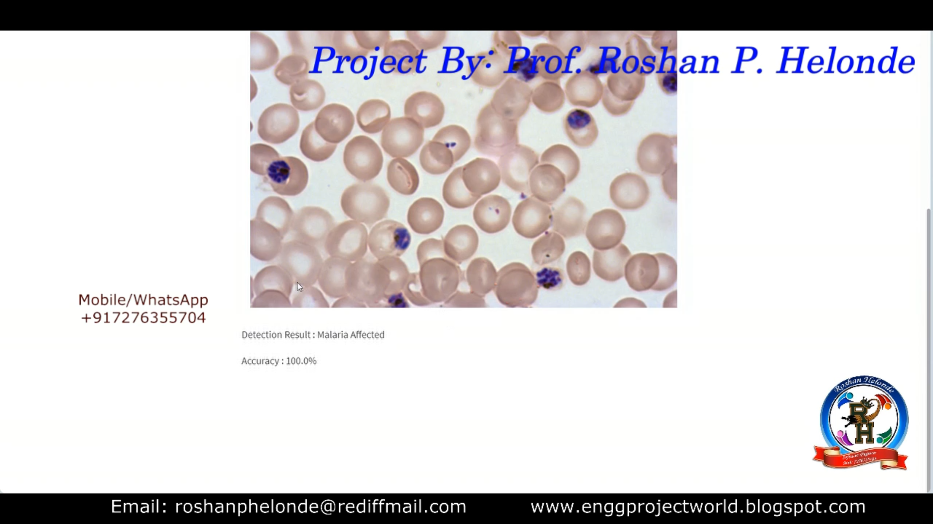
Malaria Detection Using Image Processing Python Project | Malaria Parasite Detection Using CNN | Final Year Project

 ABSTRACT

             Malaria is a life-threatening disease caused by parasites that are transmitted to people through the bites of infected female Anopheles mosquitoes. It is preventable and curable.  Malaria is a serious disease which is caused by the parasite of the genus plasmodium. It poses a global problem and warrants an automatic evaluation process because conventional microscopy which is considered the gold standard has proven to be inefficient and its results are hard to store and reproduce. In conventional microscopy the blood of a malaria infected patient is placed in a slide and is observed under a microscope. This is a time consuming and tiring process even with the involvement of an expert technician. In this study we propose a computerized diagnosis which will help in immediate detection of the disease so that proper treatment can be provided to the malaria patient. We propose the usage of image processing techniques to automate the process of parasite detection in blood samples of patients. The proposed system is robust and it is unaffected by exceptional circumstances and achieves high percentages of accuracy. This project is develop in python.

PROJECT OUTPUT


PROJECT VIDEO

Contact:
Prof. Roshan P. Helonde
Mobile: +91-7276355704
WhatsApp: +91-7276355704
Email: roshanphelonde@rediffmail.com
Share:

Diabetic Retinopathy Detection Using Image Processing Matlab Project Code | Final Year Project

 ABSTRACT

          The processing of images by performing some operations in order to get enhanced images is called as image processing. It is widely used to diagnose the eye diseases in an easy and efficient manner. Several techniques has been developed for the early detection of DR on the basis of  features  such as blood. It includes the image enhancement  processes  like  histogram  equalization  and adaptive histogram equalization for the detection of DR. The persistent  damage  caused  to  the  retina  is  termed  as  the retinopathy.  The  condition  of  diabetic  retinopathy  (DR) happens  with  those  who  have  diabetes  that  results  in progressive damage to the retina.  Due to high blood glucose levels it  leads to the  damage of small blood vessels in  the retina and this may result into swelling of the retina. ie., DR is a diabetes related eye disease which occurs when the blood vessels in the retina become swelled and leaks fluid which ultimately leads to vision loss. The DR is regarded as a serious sight threatening condition. The  main  objective of  this method  is to  detect DR (Diabetic  Retinopathy)  eye  disease  using  Image  Processing techniques. The tool used  in this method is MATLAB and it is widely used in image processing. This project proposes a method for Extraction of Blood Vessels from the medical image of human  eye-retinal  fundus  image  that  can  be  used  in ophthalmology  for  detecting  DR.  This  method  utilizes  an approach  of  Adaptive  Histogram  Equalization  using  CLAHE (Contrast  Limited Adaptive  Histogram Equalization)  algorithm with Convolutional Neural Networks algorithm implementation. The result shows that affected DR is detected in fundus image and the DR  is  not  detected  in  the  healthy  fundus  image  and upto 98%  of Accuracy can be achieved in the detection of DR Project.

PROJECT OUTPUT


PROJECT DEMO VIDEO

Contact:
Prof. Roshan P. Helonde
Mobile: +91-7276355704
WhatsApp: +917276355704
Email: roshanphelonde@rediffmail.com
Share:

Type of Skin Disease Detection Using Image Processing Matlab Project Source Code | Melanoma Disease Detection

 ABSTRACT

          Skin diseases are more common than other diseases. Skin diseases may be caused by fungal infection, bacteria, allergy, or viruses, etc. A skin disease may change texture or color of the skin. In general, skin diseases are chronic, infectious and sometimes may develop into skin cancer. The advancement of lasers and Photonics based medical technology has made it possible to diagnose the skin diseases much more quickly and accurately. But the cost of such diagnosis is still limited and very expensive. So, image processing techniques help to build automated screening system for dermatology at an initial stage. The extraction of features plays a key role in helping to classify skin diseases. Computer vision has a role in the detection of skin diseases in a variety of techniques. Due to deserts and hot weather, skin diseases are common in various country. We proposed an image processing-based method to detect skin diseases. This method takes the digital image of disease effect skin area, then use image analysis to identify the type of disease. This project is developed in matlab using image processing techniques with up to 98% Accuracy.

PROJECT OUTPUT


PROJECT VIDEO

Contact:
Prof. Roshan P. Helonde
Mobile: +91-7276355704
WhatsApp: +917276355704
Email: roshanphelonde@rediffmail.com
Share:

Diabetic Retinopathy Detection Using CNN Python Project With Source Code | Final Year Project

ABSTRACT

           Diabetic Retinopathy (DR) is a chronic health disease which requires early detection and treatment. It is important to identify DR using an intelligent system for faster prediction since manual examination and detection of the disease are unreliable and highly prone to error. Therefore, various researchers and medical experts have adopted and approached for advanced feature extraction and image classification, for early DR detection. Diabetic Retinopathy is a consequence of diabetes that affects the eyes. Damaged blood vessels in the retina, a light-sensitive tissue, are the primary cause of DR. Patients with Type 1 or Type 2 diabetes are more likely to have this condition. If the patient has a long-term case  of diabetes and  the blood sugar  level is  not regulated consistently, the odds of this  issue developing in the eye increase.  Diabetic  Retinopathy is  one  of  the most  common causes  of  blindness  in  the Western  countries. Preventing Diabetic Retinopathy has  found to be quite beneficial when people with  diabetes are  monitored regularly. This  process is  shown to be essential if Diabetic Retinopathy is discovered in its early stages due to the availability of treatment. Diabetic Retinopathy, the main cause of blindness among working-age adults, affects millions of individuals. Diabetic  Retinopathy  is  a  medical  disorder  in  which  diabetes  mellitus  causes  damage  to  the  retina.  Diabetic Retinopathy  is diagnosed  using  colored  fundus  images,  which  requires  trained clinicians  to recognize  the  presence  and importance  of  several tiny  characteristics,  making  it a  time-consuming  task.  We present  a  CNN-based  technique to  detect Diabetic Retinopathy in fundus images in this project. This project is developed in python.

PROJECT OUTPUT


PROJECT VIDEO

Contact:
Prof. Roshan P. Helonde
Mobile: +91-7276355704
WhatsApp: +91-7276355704
Email: roshanphelonde@rediffmail.com
Share:

Breast Cancer Detection Using Image Processing Matlab Project With Source Code | Final Year Project Code

ABSTRACT

        The World Health Organization's International agency for Research on Cancer in Lyon, France, estimates that more than 150000 women worldwide die of breast cancer each year. Organ chlorines are considered a possible cause for hormone-dependent cancers . Detection of early and subtle signs of breast cancer requires high-quality images and skilled mammographic interpretation. In order to detect early onset of cancers in breast screening, it is essential to have high-quality images. Radiologists reading mammograms should be trained in the recognition of the signs of early onset of, which may be subtle and may not show typical malignant features. Mammography screening programs have shown to be effective in decreasing breast cancer mortality through the detection and treatment of early onset of breast cancers. In this project we have used image processing for detection of breast cancer like Benign Cancer, Malignant Cancer and Normal Breast.

PROJECT OUTPUT


PROJECT DEMO VIDEO

Contact: 
Prof. Roshan P. Helonde 
Mobile: +91-7276355704 
WhatsApp: +91-7276355704 
Email: roshanphelonde@rediffmail.com
Share:

Skin Disease Detection Using Image Processing Matlab Project With Source Code | Melanoma Detection Final Year Project Code

ABSTRACT

         Skin cancer also known as melanoma it is one of the deadliest form of cancer if not recognized in time. Since the pigmented areas/moles of the skin can be nicely observed by simple, non-invasive visual inspection the clinical protocols of its recognition also consider several visual features. Melanoma is the deadliest form of skin cancer, which is considered one of the most common human malignancies in the world. Early detection of this disease can affect the result of the illness and improve the chance of surviving. The tremendous improvement of deep learning algorithms in image recognition tasks promises a great success for medical image analysis, in particular, melanoma classification for skin cancer diagnosis. Activation functions play an important role in the performance of deep neural networks for image recognition problems as well as medical image classification. Melanin is the pigment that discerns the color of human skin. The special cells produce melanin in the skin. If these cells are damaged or unhealthy, skin discoloration is visible. Skin pigment discoloration on cheeks is a hazardous fact as a symptom of human skin disease with a possibility of losing natural beauty. The extracted information of the skin discoloration can work as a guide to diagnosis the disease. In this project different imaging techniques like preprocessing method, segmentation and classification operations are used to analyze and extract the information of cheek’s discoloration lesion by measuring the pixel number of lesion on skin. This project is developed in matlab.

PROJECT OUTPUT


PROJECT DEMO VIDEO

Contact:
Prof. Roshan P. Helonde
Mobile: +91-7276355704
WhatsApp: +917276355704
Email: roshanphelonde@rediffmail.com
Share:

Acne Disease Detection Using CNN Python Project With Source Code | Acne Disease Detection Using Image Processing

 ABSTRACT

          Acne is a chronic skin disease occurring from inflammation of pilosebaceous units which are hair follicles under skin and their surrounding sebaceous gland (fatty gland) clog up. Currently, dermatologist has to manually mark a location of acnes on the sheet, then count to quantify and measure treatment progress. This is an unreliable and inaccurate method. Moreover, this method requires dermatologist’s excessive effort. In this project, a novel automatic acne disease detection using Image processing technique is proposed. Acne causes significant physical and psychological problems for patients such as permanent scarring, depression and anxiety from poor self-image. When you have acnes, go to see dermatologist early is the safest way to heal and prevent future permanent scars. Acne can be caused by many factors such as overactive oil glands that produce too much oil, combine with skin cells to make pores in the skin, become plugged and p-acne bacteria cause acne disease. This project is developed in python.

PROJECT OUTPUT


PROJECT DEMO VIDEO

Contact:
Prof. Roshan P. Helonde
Mobile: +91-7276355704
WhatsApp: +917276355704
Email: roshanphelonde@rediffmail.com
Share:

Total Pageviews

CONTACT US

Prof. Roshan P. Helonde
Mobile: +917276355704
WhatsApp: +917276355704
Email: roshanphelonde@rediffmail.com

Enter Project Title

Popular Projects

All Archive

Contact Form

Name

Email *

Message *