Matlab Code for Diabetic Retinopathy Detection using Convolutional Neural Network CNN

ABSTRACT
            Diabetic Retinopathy (DR) is one of the major causes of blindness in the western world. Increasing life expectancy, indulgent lifestyles and other contributing factors mean the number of people with diabetes is projected to continue rising. Regular screening of diabetic patients for DR has been shown to be a cost-effective and important aspect of their care. The accuracy and timing of this care is of significant importance to both the cost and effectiveness of treatment. If detected early enough, effective treatment of DR is available; making this a vital process. The diagnosis of diabetic retinopathy (DR) through colour fundus images requires experienced clinicians to identify the presence and significance of many small features which, along with a complex grading system, makes this a difficult and time consuming task. In this project , we propose a CNN approach to diagnosing DR from digital fundus images and accurately classifying its severity. We develop a network with CNN architecture and data augmentation which can identify Diabetic Retinopathy.
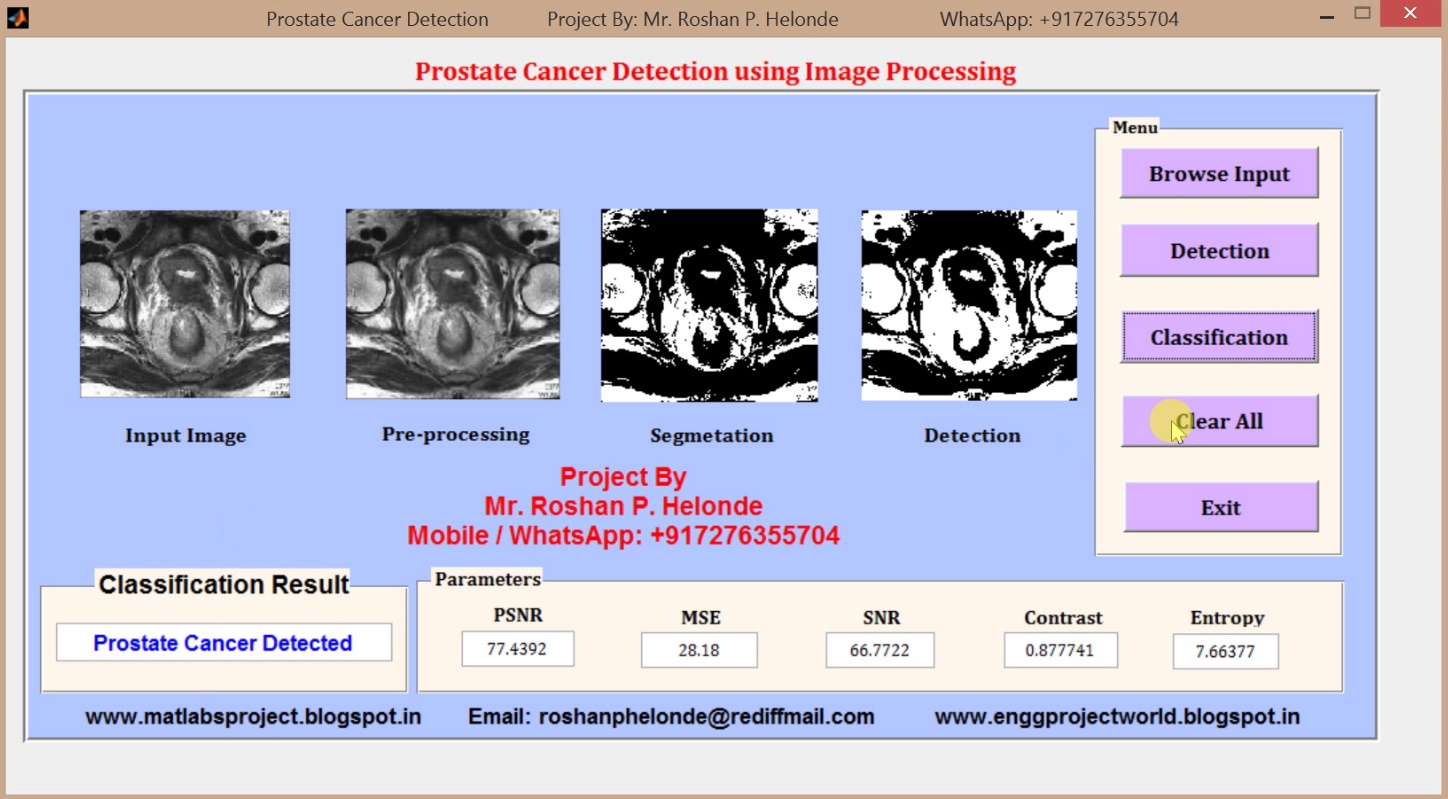
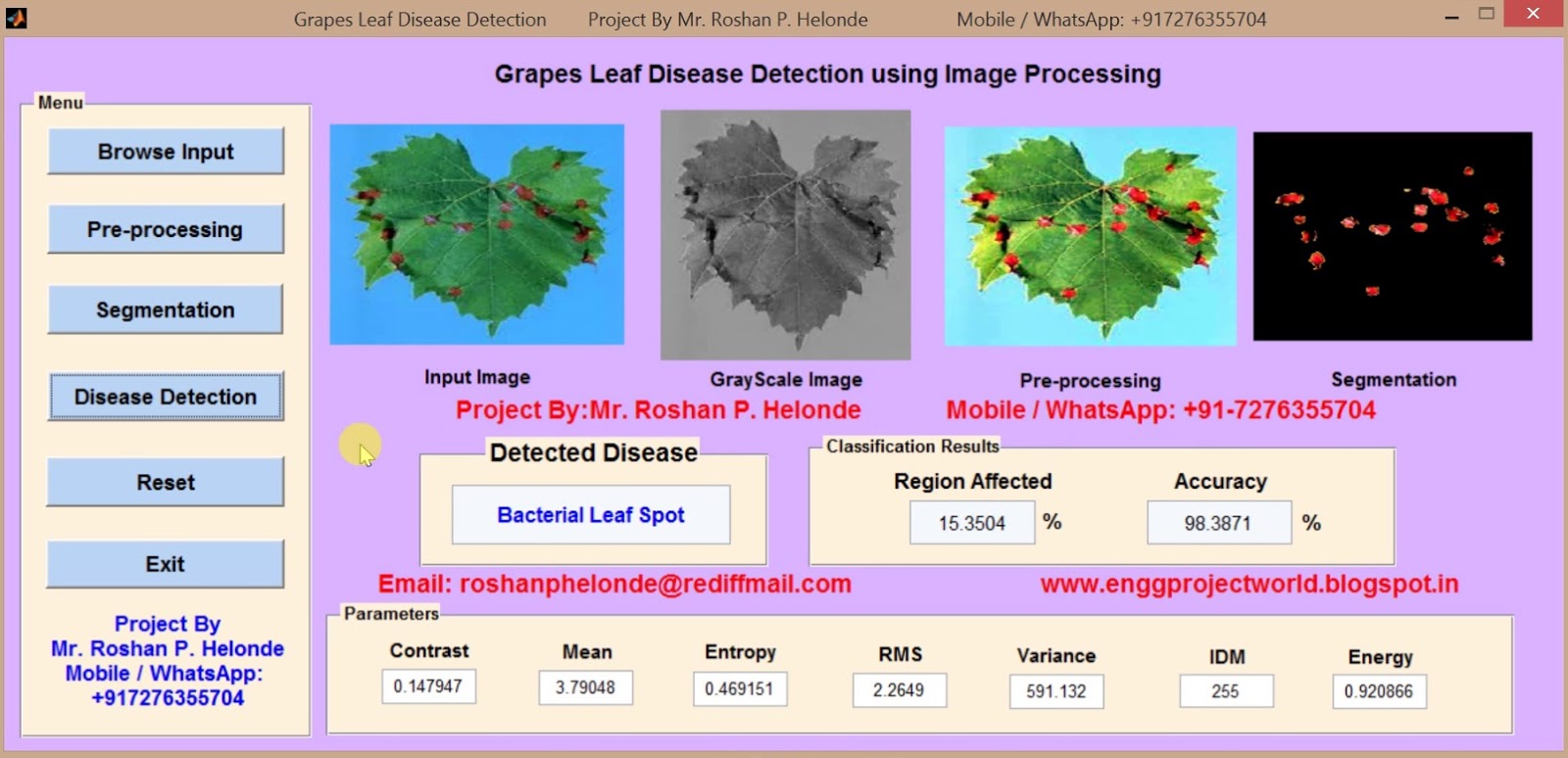
PROJECT OUTPUT

PROJECT VIDEO

Contact:
Mr. Roshan P. Helonde
Mobile: +91-7276355704
WhatsApp: +91-7276355704
Email: roshanphelonde@rediffmail.com
Share:

Matlab Code for Rice Leaf Disease Detection using Image Processing

ABSTRACT
            Agriculture is the main backbone for most of the developing/developed countries; agriculture production itself is the main feed for ever growing populations and it is the major source of income for the rural people/farmers especially in India. In India farmers are called “the backbone of India”. The main aim of the proposed system is to detect and classify the diseases in Rice leafs. Rice Diseases Classification comprises of two steps: first one is Detection, Extraction and Segmentation of diseases. Secondly, Feature extraction, Classification level of disease by using Support Vector Machine (SVM) classifiers respectively. The proposed system has been experimentally tested for our own dataset and results achieved are encouraging.

PROJECT OUTPUT

PROJECT VIDEO

Contact:
Mr. Roshan P. Helonde
Mobile: +91-7276355704
WhatsApp: +91-7276355704
Email: roshanphelonde@rediffmail.com
Share:

Matlab code for Blood Group Detection using Neural Network

ABSTRACT
           Determining of blood types is very important during emergency situation before administering a blood transfusion. Presently, these tests are performed manually by technicians, which can lead to human errors. Determination of the blood types in a short period of time and without human errors is very much essential. A method is developed based on processing of images acquired during the slide test. The image processing techniques such as Pre-processing, Segmentation, Thresholding, Morphological operations and Neural Network  are used. The images of the slide test are obtained from the pathological laboratory are processed and the occurrence of agglutination are evaluated. Thus the developed automated method determines the blood type using image processing techniques. The developed method is useful in emergency situation to determine the blood group without human error. The slide test consists of the mixture of one drop of blood and one drop of reagent, being the result interpreted according to the occurrence or not of agglutination. The combination of the occurrence and non occurrence of the agglutination determines the blood type of the patient. 

PROJECT OUTPUT

PROJECT VIDEO
Contact:
Mr. Roshan P. Helonde
Mobile: +91-7276355704
WhatsApp: +91-7276355704
Email: roshanphelonde@rediffmail.com

Share:

Hand Bone Fracture Detection using Image Processing Matlab Project with source Code

ABSTRACT
            Analysis of medical images plays a very important role in clinical decision making. For a long time it has required extensive involvement of a human expert. However, recent progress in data mining techniques, especially in machine learning, allows for creating decision models and support systems that help to automatise this task and provide clinicians with patient-specific therapeutic and diagnostic suggestions. In this project, we describe a study aimed at building a decision model (a classifier) that would predict the type of treatment (surgical vs. non-surgical) for patients with bone fractures based on their X-ray images. We consider two types of features extracted from images (structural and textural) and used them to construct multiple classifiers that are later evaluated in a computational experiment. Structural features are computed by applying the Hough transform, while textural information is obtained from Gray-level occurrence matrix. In research reported by other authors structural and textural features were typically considered separately. Our findings show that while structural features have better predictive capabilities, they can benefit from combining them with textural ones.

PROJECT OUTPUT

PROJECT VIDEO

Contact:
Mr. Roshan P. Helonde
Mobile: +91-7276355704
WhatsApp: +91-7276355704
Email: roshanphelonde@rediffmail.com
Share:

Breast Cancer Detection using Neural Network Matlab Project Code

ABSTRACT
            The World Health Organization's International agency for Research on Cancer in Lyon, France, estimates that more than 150 000 women worldwide die of breast cancer each year. The breast cancer is one among the top three cancers in American women. In United States, the American Cancer Society estimates that, 215 990 new cases of breast carcinoma has been diagnosed, in 2004. It is the leading cause of death due to cancer in women under the age of 65 . In India, breast cancer accounts for 23% of all the female cancers followed by cervical cancers (17.5%) in metropolitan cities such as Mumbai, Calcutta, and Bangalore. However, cervical cancer is still number one in rural India. Although the incidence is lower in India than in the developed countries, the burden of breast cancer in India is alarming. Organ chlorines are considered a possible cause for hormone-dependent cancers . Detection of early and subtle signs of breast cancer requires high-quality images and skilled mammographic interpretation. In order to detect early onset of cancers in breast screening, it is essential to have high-quality images. Radiologists reading mammograms should be trained in the recognition of the signs of early onset of, which may be subtle and may not show typical malignant features. Mammography screening programs have shown to be effective in decreasing breast cancer mortality through the detection and treatment of early onset of breast cancers.
          Emotional disturbances are known to occur in patient's suffering from malignant diseases even after treatment. This is mainly because of a fear of death, which modifies Quality Of Life (QOL). Desai et al.,reported an immuno histo chemical analysis of steroid receptor status in 798 cases of breast tumors encountered in Indian patients, suggests that breast cancer seen in the Indian population may be biologically different from that encountered in western practice. Most imaging studies and biopsies of the breast are conducted using mammography or ultrasound, in some cases, magnetic resonance (MR) imaging . Although by now some progress has been achieved, there are still remaining challenges and directions for future research such as developing better enhancement and segmentation algorithms. 

PROJECT OUTPUT

PROJECT VIDEO

Contact: 
Mr. Roshan P. Helonde 
Mobile: +91-7276355704 
WhatsApp: +91-7276355704 
Email: roshanphelonde@rediffmail.com
Share:

Audio Steganography (Hiding Secrete Information in Audio) Matlab Project Code

ABSTRACT
           Today’s large demand of internet applications requires data to be transmitted in a secure manner. Data transmission in public communication system is not secure because of interception and improper manipulation by eavesdropper. So the attractive solution for this problem is Steganography, which is the art and science of writing hidden messages in such a way that no one, apart from the sender and intend recipient, suspects the existence of the message, a form of security through obscurity. Audio steganography is the scheme of hiding the existence of secret information by concealing it into another medium such as audio file. The steganography application hides different types of data within a cover file. The resulting stego also contains hidden information, although it is virtually identical to the cover file.

PROJECT OUTPUT

PROJECT VIDEO

Contact:
Mr. Roshan P. Helonde
Mobile: +91-7276355704
WhatsApp: +91-7276355704
Email: roshanphelonde@rediffmail.com
Share:

Matlab code for Brain Tumor Detection using Convolutional Neural Network (CNN)

ABSTRACT
           Brain tumor identification is really challenging task in early stages of life. But now it became advanced with various machine learning algorithms. Now a day’s issue of brain tumor automatic identification is of great interest. In Order to detect the brain tumor of a patient we consider the data of patients like MRI images of a patient’s brain. Here our problem is to identify whether tumor is present in patients brain or not. It is very important to detect the tumors at starting level for a healthy life of a patient. There are many literatures on detecting these kinds of brain tumors and improving the detection accuracies. In this project, we Estimate the brain tumor severity using Convolutional Neural Network algorithm which gives us accurate results.

PROJECT OUTPUT

PROJECT VIDEO

Contact:
Mr. Roshan P. Helonde
Mobile: +91-7276355704
WhatsApp: +91-7276355704
Email: roshanphelonde@rediffmail.com
Share:

Image Fusion using Wavelet Transform Matlab Source code

ABSTRACT
            Different medical imaging techniques such as X-rays, computed tomography (CT), magnetic resonance imaging (MRI), and positron emission tomography (PET) provide different perspectives for the human body that are important in the physical disorders or diagnosis of diseases .To derive useful information from multimodality medical image data medical image fusion has been used. In the medical field different radiometric scanning techniques can be used to evaluate and examine the inner parts of the body. The idea is to improve the image content by fusing images like computer tomography (CT) and magnetic resonance imaging (MRI) images, so as to provide as much details as possible for the sake of diagnosis. The objective of image fusion is to merge information from multiple images of the same image. The resultant image after image fusion is more suitable for human and machine perception and further helpful for image-processing tasks such as segmentation, feature extraction and object recognition. This paper mainly presents image fusion using wavelet method for multispectral data and high-resolution data conveniently, quickly and accurately in MATLAB. Wavelet toolbox with abundant functions, provide a quick and convenient platform to improve image visibility. The work covers the selection of wavelet function, the use of wavelet based fusion algorithms on CT and MRI medical images, implementation of fusion rules and the fusion image quality evaluation. Matlab Results show that effectiveness of Image Fusion with Wavelet Transform on preserving the feature information for the test images.

PROJECT OUTPUT

PROJECT VIDEO

Contact:
Mr. Roshan P. Helonde
Mobile: +91-7276355704
WhatsApp: +91-7276355704
Email: roshanphelonde@rediffmail.com
Share:

Handwritten Digit Recognition using Neural Network Matlab Source code

ABSTRACT
           Handwriting recognition is one of the compelling research works going on because every individual in this world has their own style of writing. It is the capability of the computer to identify and understand handwritten digits automatically. Because of the progress in the field of science and technology, everything is being digitalised to reduce human effort. Hence, there comes a need for handwritten digit recognition in many real-time applications. Many Machine Learning and Deep Learning Algorithms are developed which can be used for this digit classification. This project performs Digit Recognition and the analysis of accuracy of algorithms Neural Network.

PROJECT OUTPUT

PROJECT VIDEO

Contact:
Mr. Roshan P. Helonde
Mobile: +91-7276355704
WhatsApp: +91-7276355704
Email: roshanphelonde@rediffmail.com
Share:

Image Fusion using Wavelet Transform and PCA Matlab Source code

ABSTRACT
            Different medical imaging techniques such as X-rays, computed tomography (CT), magnetic resonance imaging (MRI) provide different perspectives for the human body that are important in the physical disorders or diagnosis of diseases .To derive useful information from multimodality medical image data medical image fusion has been used. In the medical field different radiometric scanning techniques can be used to evaluate and examine the inner parts of the body. The idea is to improve the image content by fusing images like computer tomography (CT) and magnetic resonance imaging (MRI) images, so as to provide as much details as possible for the sake of diagnosis. The objective of image fusion is to merge information from multiple images of the same image. The resultant image after image fusion is more suitable for human and machine perception and further helpful for image-processing tasks such as segmentation, feature extraction and object recognition. This project mainly presents image fusion using wavelet method for multispectral data and high-resolution data conveniently, quickly and accurately in MATLAB. Wavelet toolbox with abundant functions, provide a quick and convenient platform to improve image visibility. The work covers the selection of wavelet function, the use of wavelet based fusion algorithms on CT and MRI medical images, implementation of fusion rules and the fusion image quality evaluation. Matlab Results show that effectiveness of Image Fusion with Wavelet Transform on preserving the feature information for the test images.

PROJECT OUTPUT

PROJECT VIDEO

Contact:
Mr. Roshan P. Helonde
Mobile: +91-7276355704
WhatsApp: +91-7276355704
Email: roshanphelonde@rediffmail.com
Share:

Image Enhancement using Histogram Equalization Matlab Source code

ABSTRACT
             Digital image enhancement is one of the most important image processing technology which is necessary to improve the visual appearance of the image or to provide a better transform representation for future automated image processing such as image analysis, detection, segmentation and recognition. Many images have very low dynamic range of the intensity values due to insufficient illumination and therefore need to be processed before being displayed. Large number of techniques have focused on the enhancement of gray level images in the spatial domain. These methods include histogram equalization, gamma correction, high pass filtering, low pass filtering, homomorphic filtering, etc. Image enhancement techniques are of particular interest in photography, satellite imagery, medical applications and display devices. Producing visually natural is required for many important areas such as vision, remote sensing, dynamic scene analysis, autonomous navigation, and biomedical image analysis.

PROJECT OUTPUT

PROJECT VIDEO

Contact:
Mr. Roshan P. Helonde
Mobile: +91-7276355704
WhatsApp: +91-7276355704
Email: roshanphelonde@rediffmail.com
Share:

Prostate Cancer Detection using Image Processing Matlab Source Code

ABSTRACT
          This project gives an overview of the method of detecting prostate cancer by associating Region of interest segmentation method with Support Vector Machine. Prostate cancer is commonly prevalent carcinoma detected in most of the male population. A diagnosis of prostate cancer was complicated due to unclear symptoms and involves many procedures. One of these procedures involves the study of prostate tissue biopsy to find cancer affected region. However, no boundary specified region was considered for further studies. Recent developmental techniques in the medical imaging field, especially in SVM, have paved the way for prostate carcinoma detection. The MRI image of the prostate gland is pre-processed to reduce noise effects and Region of interest is obtained with the svm and segmentation is done. The core idea of this project is to assume that every region of prostate tissue could be related to malignant or unnatural tissues.

PROJECT OUTPUT

PROJECT VIDEO

Contact:
Mr. Roshan P. Helonde
Mobile: +91-7276355704
WhatsApp: +91-7276355704
Email: roshanphelonde@rediffmail.com
Share:

Grape Leaf Disease Detection using Image Processing Matlab Source code

ABSTRACT
             Identification of the grape leaf disease is the main goal to prevent the losses and quality of agricultural product. In India grape fruit crop is widely grown. So disease detection and classification of grape leaf is very critical for sustainable agriculture. It’s not possible to farmer, to monitor continuously the grape disease manually. It requires the excessive processing time, tremendous amount of work, and some expertise in the grape leaf diseases. To detect and classify the grape disease we need fast automatic process so we use SVM classifier technique. This project presents mainly five stages, viz image acquisition, pre-processing, segmentation GLCM feature extraction and SVM classification. This project is proposed to benefit in the detection and classification of grape leaf disease using support vector machine (SVM) classifier.

PROJECT OUTPUT

PROJECT VIDEO

Contact:
Mr. Roshan P. Helonde
Mobile: +91-7276355704
WhatsApp: +91-7276355704
Email: roshanphelonde@rediffmail.com
Share:

Total Pageviews

CONTACT US

Prof. Roshan P. Helonde
Mobile: +917276355704
WhatsApp: +917276355704
Email: roshanphelonde@rediffmail.com

Enter Project Title

Popular Projects

All Archive

Contact Form

Name

Email *

Message *