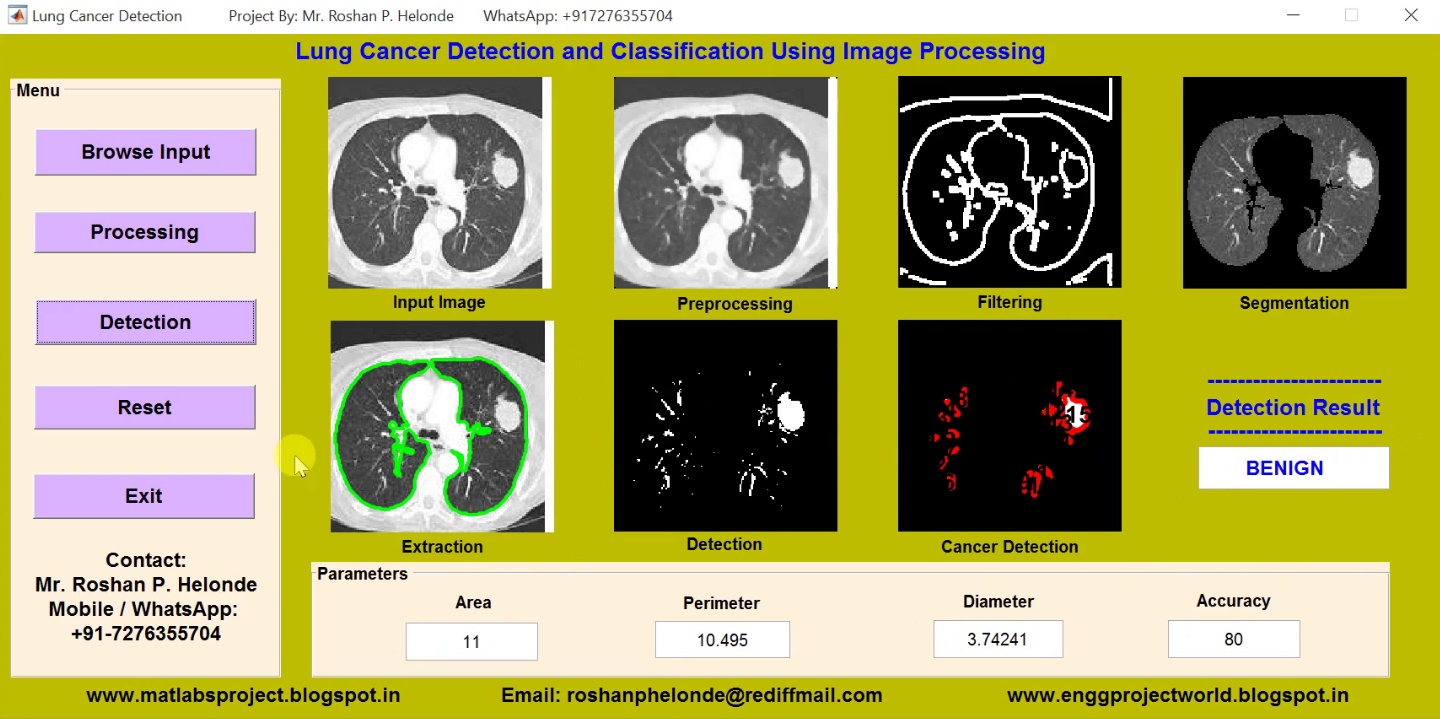
Gender Recognition System from Speech Signal Matlab Project with Source Code IEEE Project

ABSTRACT
             Signal is a physical quantity that varies with respect to the independent variable like time, space, etc. Signal values can be represented in zero’s and one’s. Processing of digital signal by using digital computer is called as Digital Signal Processing. According to Webster’s dictionary, speech is the expression or communication throughout in speakers. Speech is the most important thing to express our thoughts. Speech signal is used to communicate among people. It not only consists of the information but also carries the information regarding the particular speaker. From which the speaker is male or female can be recognised. The meaning of Gender Recognition (GR) is recognising the gender of the person whether the speaker is male or female. The Information about gender, age, ethnicity, and emotional state are the important ingredients that give rich behavioural information. Such information can be obtained from the speech signal. In this project, an unknown speaker is compared to a database of some known speakers. The best matching system is taken as the recognition decision. From the Recognition decision we conclude whether the given voice sample is generated by a male or female.

PROJECT OUTPUT

PROJECT VIDEO

Contact:
Mr. Roshan P. Helonde
Mobile: +91-7276355704
WhatsApp: +91-7276355704
Email: roshanphelonde@rediffmail.com
Share:

Shape Recognition Using Image Processing Matlab Project with Source Code IEEE Project

ABSTRACT
                    Doing image processing and especially blob analysis it is often required to check some objects' shape and depending on it perform further processing of a particular object or not. For example, some applications may require finding only circles from all the detected objects, or quadrilaterals, rectangles, etc. Human vision seems to make use of many sources of information to detect and recognise an object in a scene. At the lowest level of object recognition, researchers agree that edge and region information are utilised to extract a “perceptual unit” in the scene. Some of the possible invariant features are recognised and additional signal properties (texture or appearance) are sent to help in making the decision as to whether a point belongs to an object or not. In many cases, boundary shape information, such as the rectangular shapes of vehicles in aerial imagery, seems to play a crucial role. Local features such as the eyes in a human face are sometimes useful. These features provide strong clues for recognition, and often they are invariant to many scene variables.The study of shapes is a recurring theme in computer vision. For example, shape is one of the main sources of information that can be used for object recognition. In medical image analysis, geometrical models of anatomical structures play an important role in automatic tissue segmentation. The shape of an organ can also be used to diagnose diseases. In a completely different setting, shape plays an important role in the perception of optical illusions (we tend to see particular shapes) and this can be used to explain how our visual system interprets the ambiguous and incomplete information available in an image. Characterizing the shape of a specific rigid object is not a particularly hard problem, although using the shape information to solve perceptual tasks is not easy.

PROJECT OUTPUT

PROJECT VIDEO

Contact:
Mr. Roshan P. Helonde
Mobile: +91-7276355704
WhatsApp: +91-7276355704
Email: roshanphelonde@rediffmail.com
Share:

Matlab Code for Glaucoma Detection Using Image Processing

ABSTRACT
                Computational techniques have great impact in the field of Medicine and Biology. These techniques help the medical practitioners to diagnose any abnormality in advance and provide fruitful treatment. Retinal image analysis has been an ongoing area of research. Automated retinal image analysis aid the ophthalmologists in detecting abnormalities in the retinal structures namely optic disc, blood vessels, thus diagnosing sight threatening retinal diseases such as Glaucoma and Retinopathy. Glaucoma is the major cause of blindness in working population. Glaucoma is characterised by increased intra-ocular pressure inside the eye leading to changes in the optic disc and optic nerve. It does not reveal its symptoms until later stage. Hence, regular screening of the patients is required to identify the disease, thus demanding high labor, time and expertise. Thus, computational techniques are sought for their analysis.
                 In this project, identification of Glaucoma is carried out through computational techniques namely image processing. As the changes in the profile of optic disc act as a biomarker for the onset of the disease, optic disc is segmented through image processing techniques. Optic disc is the brightest part portrayed as oval structure in the retinal fundus image. It encompasses optic cup, which is the brightest central part, optic rim, the surrounding pale part and the blood vessels. All these structures are segmented and their properties are elicited. Then, properties of the disc, cup and blood vessels within optic disc are mined to design a learning model for prediction of Glaucoma.

PROJECT OUTPUT

PROJECT VIDEO

Contact:
Mr. Roshan P. Helonde
Mobile: +91-7276355704
WhatsApp: +91-7276355704
Email: roshanphelonde@rediffmail.com
Share:

Matlab Code for Lung Cancer Detection Using Image Processing

ABSTRACT
        Lung cancer prevalence is one of the highest of cancers, at 18 %. One of the first steps in lung cancer diagnosis is sampling of lung tissues or biopsy. These tissue samples are then microscopically analyzed. This procedure is taken once imaging tests indicate the presence of cancer cells in the chest. Lung cancer diagnosis using lung images. One of them is that doctor still relies on subjective visual observation. A medical specialist must do thorough observation and accurate analysis in detecting lung cancer in patients. Hence, there is need for a system that is capable for detecting lung cancer automatically from microscopic images of biopsy. This method will improve the accuracy and efficiency for lung cancer detection. The aim of this research is to design a lung cancer detection system based on analysis of microscopic image of biopsy using digital image processing. Microscopic images of biopsy are feature extracted and classified using support vector machine. This method is implemented to detection of lung cancer of lung samples.

PROJECT OUTPUT

PROJECT VIDEO

Contact:
Mr. Roshan P. Helonde
Mobile: +91-7276355704
WhatsApp: +91-7276355704
Email: roshanphelonde@rediffmail.com

Share:

Matlab Code for Fake Currency Recognition Using Image Processing IEEE Project

ABSTRACT
                  The Reserve Bank is the one which issue bank notes in India. Reserve Bank, changes the design of bank notes from time to time. Reserve bank uses several techniques to detect fake currency. Common people faces many problems for the fake currency circulation and also difficult to detect fake currency, suppose that a common people went to a bank to deposit money in bank but only to see that some of the notes are fake, in this case he has to take the blame. As banks will not help that person. Some of the effects that fake currency has on society include a reduction in the value of real money; and inflation due to more fake currency getting circulated in the society or market which disturbs our economy and growth - an some illegal authorities an artificial increase in the money supply,a decrease in the acceptability of paper money and losses. Our aim is to help common man to recognize currency. Proposed system is based on image processing and makes the process automatic and robust. Shape information are used in our algorithm. Original Note Detection Systems are present in banks but are very costly. We are developing an image processing algorithm which will extract the currency features and compare it with features of original note image. This system is cheaper and can provide accuracy on the basics of visual contents of note.

PROJECT OUTPUT

PROJECT VIDEO

Contact:
Mr. Roshan P. Helonde
Mobile: +91-7276355704
WhatsApp: +91-7276355704
Email: roshanphelonde@rediffmail.com
Share:

Matlab Code for Brain Tumor Detection and Classification Using Image Processing

ABSTRACT
          Brain is the kernel part of the body. Brain has a very complex structure. Brain is hidden from direct view by the protective skull. This skull gives brain protection from injuries as well as it hinders the study of its function in both health and disease. But brain can be affected by a problem which cause change in its normal structure and its normal behavior .This problem is known as brain tumor. Brain tumor causes the abnormal growth of the cells in the brain. The cells which supplies the brain in the arteries are tightly bound together thereby routine laboratory test are inadequate to analyze the chemistry of brain. Brain tumor diagnosis is a very crucial task. This system provides an efficient and fast way for diagnosis of the brain tumor. Proposed system consists of multiple phases. First phase consists of texture feature extraction from brain MR images. Second phase classify brain images on the bases of these texture feature using ensemble base classifier. After classification tumor region is extracted from those images which are classified as malignant or benign. Segmentation consists of tumor extraction phases. Quantitative results show that our proposed system performed very efficiently and accurately.

PROJECT OUTPUT

PROJECT VIDEO

Contact:
Mr. Roshan P. Helonde
Mobile: +91-7276355704
WhatsApp: +91-7276355704
Email: roshanphelonde@rediffmail.com
Share:

Matlab Code for OMR Answer Sheet Evaluation

ABSTRACT
           This project aims to develop Image processing based Optical Mark Recognition sheet scanning system. Today we find that lot of competitive exams are been conducted as entrance exams. These exams consists of MCQs. The students have to fill the right box or circle for the appropriate answer to the respective questions. During the inspection or examining phase normally a stencil is provided to the examiner to determine the right answer to the questions. This is a manual process and a lot of errors can occur in the manual process such as counting mistake and many more. To avoid this mistakes OMR system is used. In this system OMR answer sheet will be scanned and the scanned image of the answer sheet will be given as input to the software system. Using Image processing we will find the answers marked to each of the questions. Summation of the marks & displaying of total marks will be also implemented. The implementation is done using Matlab
        In today’s modern world of technology when everything is computerized, the Evaluation exercise of examining and assessing the educational system has become absolute necessity. Today, more emphasis is on objective exam which is preferred to analyze scores of the students since it is simple and requires less time in the examining objective answer-sheet as compared to the subjective answer-sheet. This project proposes a new technique for generating scores of multiple-choice tests which are done by developing a technique that has software based approach with computer & scanner which is simple, efficient & reliable to all with minimal cost. Its main benefit to work with all available scanners, In addition no special paper & colour required for printing for marksheet. To recognize & allot scores to the answer marked by of the student’s.

PROJECT OUTPUT

PROJECT VIDEO

Contact:
Mr. Roshan P. Helonde
Mobile: +91-7276355704
WhatsApp: +91-7276355704
Email: roshanphelonde@rediffmail.com
Share:

Matlab Code for Real Time Video Surveillance System

ABSTRACT
             A key technology to fight against terrorism and crime for public safety moving object detection and tracking is become very popular and one of the challenging research topic in various security areas of computer vision and video surveillance applications. Due to increase in criminal and terrorist activities, general social problems, providing the security to citizens, private places, public places has become more important. Therefore watch for 24*7 is required in area of automatic monitoring. The video surveillance system does this job as accurately as possible. The video surveillance system described here is interfacing of camera and alarm system with the computer. Here the video is taken from camera and the unwanted entities are identified using MATLAB. Security, Surveillance, General identity verification, Criminal justice systems, Image database investigations, Smart Card applications are important issues in today’s world. The recent acts of terrorism require urgent need for efficient surveillance. Now a day surveillance systems use digital video recording (DVR) cameras which play host to multiple channels and the major drawbacks with this model is that it requires continuous manual monitoring and cost of manual labour. It is virtually impossible to search through recordings for important events in the past since that would require a playback of the entire duration of video footage. Hence, there is a need for an automated system for video surveillance. 

PROJECT OUTPUT

PROJECT VIDEO

Contact:
Mr. Roshan P. Helonde
Mobile: +91-7276355704
WhatsApp: +91-7276355704
Email: roshanphelonde@rediffmail.com
Share:

Matlab Code for Skin Cancer Detection Using Image Processing

ABSTRACT
         Skin cancer – also known as malignant melanoma – is one of the deadliest form of cancer if not recognized in time. Since the pigmented areas/moles of the skin can be nicely observed by simple, non-invasive visual inspection (e.g. by a dermatoscope), the clinical protocols of its recognition also consider several visual features. Melanoma is the deadliest form of skin cancer, which is considered one of the most common human malignancies in the world. Early detection of this disease can affect the result of the illness and improve the chance of surviving. The tremendous improvement of deep learning algorithms in image recognition tasks promises a great success for medical image analysis, in particular, melanoma classification for skin cancer diagnosis. Activation functions play an important role in the performance of deep neural networks for image recognition problems as well as medical image classification. Melanin is the pigment that discerns the color of human skin. The special cells produce melanin in the skin. If these cells are damaged or unhealthy, skin discoloration is visible. Skin pigment discoloration on cheeks is a hazardous fact as a symptom of human skin disease with a possibility of losing natural beauty. The extracted information of the skin discoloration can work as a guide to diagnosis the disease. In this research, different imaging techniques like watershed method, edge detection and morphological operations are used to analyze and extract the information of cheek’s discoloration lesion by measuring the pixel number of lesion on skin. The image analyzing results are visually examined by the skin specialist.

PROJECT OUTPUT

PROJECT VIDEO

Contact:
Mr. Roshan P. Helonde
Mobile: +91-7276355704
WhatsApp: +91-7276355704
Email: roshanphelonde@rediffmail.com

Share:

Heart Disease Detection using Matlab Project with Source Code

ABSTRACT
            Modern day lifestyle and our ignorance towards health have put the most vital organ of our body Heart at great risk. India today is witnessing a lot many young people suffering from heart diseases which even lead to untimely demise. Most common heart abnormality includes arrhythmia which is nothing but irregular beating of heart. Going by the trend/statistics, middle aged people (30-45yrs) are at great risk because of high stress in both personal and professional lives. This necessitates the need for a system which can not only detect any anomaly in functioning of our heart but warns us against any threat. Our project is based on developing such a system that can give us prior information about the upcoming threat or the heart disease which we are prone to. Cardiac arrhythmia is a major kind of abnormal heart activity. An arrhythmia is a problem with the heartbeat rate or rhythm of the heartbeat. For the period of an arrhythmia, the heart may beat too fast or too slow, or with an irregular rhythm. Fast heartbeat is said to be tachycardia whereas slow is called Bradycardia. Classification of cardiac arrhythmia is a difficult task. One of the ways to detect cardiac arrhythmia is to use electrocardiogram (ECG) signals. The ECG is the most important biomedical-signal used by cardiologists for diagnostic purposes. The ECG signal provides all the required information about the electrical activity of the heart. The early detection of the cardiac arrhythmias can save lives and enhance the quality of living through appreciates treatment.

PROJECT OUTPUT

PROJECT VIDEO

Contact:
Mr. Roshan P. Helonde
Mobile: +91-7276355704
WhatsApp: +91-7276355704
Email: roshanphelonde@rediffmail.com
Share:

Brain Tumor Detection Using Neural Network Matlab Code

ABSTRACT
          The imaging plays a central role in the diagnosis of brain tumors. An efficient algorithm is proposed in this project for brain tumor detection based on digital image segmentation. Brain tumor may be considered among the most difficult tumors to treat, as it involves the organ which is not only in control of the body. We proposed an Artificial Neural Network Approach for Brain Tumor Detection, which gave the edge pattern and segment of brain and brain tumor itself. The segmentation of brain tumors in magnetic resonance images is a challenging and difficult task because of the variety of their possible shapes, locations, image intensities. In this project it is intended to summarize and compare the methods of automatic detection of brain tumor through Magnetic Resonance Image used in different stages of Computer Aided Detection System. Brain Image classification techniques are studied. Existing methods are classically divided into region based and contour based methods. These are usually dedicated to full enhanced tumors or specific types of tumors. The amount of resources required to describe large set of data is simplified and selected in for tissue segmentation. In this project image segmentation techniques were applied on input images in order to detect brain tumors. Also in this project a Neural Network model that is based on machine learning with image and data analysis and manipulation techniques is proposed to carry out an automated brain tumor classification.

PROJECT OUTPUT

PROJECT VIDEO

Contact:
Mr. Roshan P. Helonde
Mobile: +91-7276355704
WhatsApp: +91-7276355704
Email: roshanphelonde@rediffmail.com
Share:

Total Pageviews

CONTACT US

Prof. Roshan P. Helonde
Mobile: +917276355704
WhatsApp: +917276355704
Email: roshanphelonde@rediffmail.com

Enter Project Title

Popular Projects

All Archive

Contact Form

Name

Email *

Message *