Agriculture is the main backbone for most of the developing/developed countries; agriculture production itself is the main feed for ever growing populations and it is the major source of income for the rural people/farmers especially in India. In India farmers are called “the backbone of India”. The main aim of the proposed system is to detect, classify the diseases and suggest pesticide to recover from disease in paddy leafs. Paddy leaf Diseases Classification done using Convolutional Neural Network (CNN) classifiers and then suggesting pesticide respectively. The proposed system has been experimentally tested for our own dataset and results achieved are encouraging.
During the last decade information security has become the major issue. The encrypting and decrypting of the data has been widely investigated because the demand for the better encryption and decryption of the data is gradually increased for getting the better security for the communication between the devices more privately. The cryptography play a major role for the fulfillment for this demand. The purpose of this project is to provide the better as well as more secure communication system by enhancing the strength of Advance Encryption Standard (AES) algorithm. AES algorithm was known for providing the best security without any limitations.
Leukemia Blood cancer is the most prevalent and it is very much dangerous among all type of cancers. Early detection of blood cancer has the potential to reduce mortality and morbidity. There are many diagnostic technologies and tests to diagnose blood cancer. However many of these tests are extremely complex and subjective and depend heavily on the experience of the technician. To obviate these problems, image processing techniques is use in this study as promising modalities for detection of Leukemia blood cancer. The accuracy rate of the diagnosis of blood cancer by using image processing will be yield a slightly higher rate of accuracy then other traditional methods and will reduce the effort and time. We first discuss the preliminary of cell biology required to proceed to implement our proposed method. This project presents a new automated approach for blood Cancer detection and analysis from a given photograph of patient’s cancer affected blood sample. The proposed method is using image improvement, image segmentation for segmenting the different cells of blood, edge detection for detecting the boundary, size, and shape of the cells and finally clustering for final decision of blood cancer based on the number of different cells.
The World Health Organization's International agency for Research on Cancer in Lyon, France, estimates that more than 150 000 women worldwide die of breast cancer each year. Organ chlorines are considered a possible cause for hormone-dependent cancers . Detection of early and subtle signs of breast cancer requires high-quality images and skilled mammographic interpretation. In order to detect early onset of cancers in breast screening, it is essential to have high-quality images. Radiologists reading mammograms should be trained in the recognition of the signs of early onset of, which may be subtle and may not show typical malignant features. Mammography screening programs have shown to be effective in decreasing breast cancer mortality through the detection and treatment of early onset of breast cancers.
Digital Image watermarking is a technology for embedding various types of information in digital content. In general, information for protecting copyrights and proving the validity of data is embedded as a watermark. A digital watermark is a digital signal or pattern inserted into digital content. The digital content could be a still image, an audio clip, a video clip, a text document, or some form of digital data that the creator or owner would like to protect. The main purpose of the watermark is to identify who the owner of the digital data is, but it can also identify the intended recipient. The Image watermarking is most popular method for copyright protection by discrete wavelet transform which performs two level decomposition of original cover image and watermark image is embedded in lowest level sub band of cover image.
Diabetic Retinopathy (DR) is one of the major causes of blindness in the western world. Increasing life expectancy, indulgent lifestyles and other contributing factors mean the number of people with diabetes is projected to continue rising. Regular screening of diabetic patients for DR has been shown to be a cost-effective and important aspect of their care. The accuracy and timing of this care is of significant importance to both the cost and effectiveness of treatment. If detected early enough, effective treatment of DR is available; making this a vital process. The diagnosis of diabetic retinopathy (DR) through colour fundus images requires experienced clinicians to identify the presence and significance of many small features which, along with a complex grading system, makes this a difficult and time consuming task. In this project , we propose a CNN approach to diagnosing DR from digital fundus images and accurately classifying its severity. We develop a network with CNN architecture and data augmentation which can identify Diabetic Retinopathy.
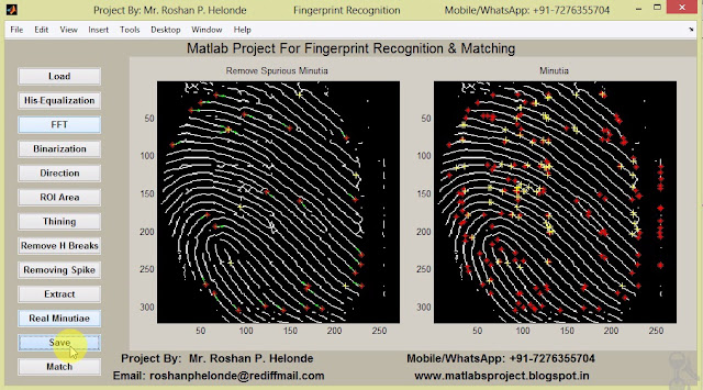
The popular Biometric used to authenticate a person is Fingerprint which is unique and permanent throughout a person’s life. A minutia matching is widely used for fingerprint recognition and can be classified as ridge ending and ridge bifurcation. In this project we projected Fingerprint Recognition using Minutia Score Matching method (FRMSM). For Fingerprint thinning, the Block Filter is used, which scans the image at the boundary to preserves the quality of the image and extract the minutiae from the thinned image. Fingerprint is a very vital concept in making us completely unique and can not be altered. It is necessary to recognize fingerprint in proper manner. Here we are trying to recognize the fingerprint image samples by using minute extraction and minute matching techniques. In minute extraction it counts the crossing numbers and from the count it will be classified as normal ridge pixel, termination point and bifurcation point. Then the input finger print data is compared with the template data. This is called as minute matching.
The detection and classification of plant diseases are the crucial factors in plant production and the reduction of losses in crop yield. This project proposes an approach for plant leaf disease detection and classification on plants using image processing. The plant disease diagnosis is restricted by person’s visual capabilities as it is microscopic in nature. Due to optical nature of plant monitoring task, computer visualization methods are adopted in plant disease recognition. The aim is to detect the symptoms of the disease occurred in leaves in an accurate way. Once the captured image is pre-processed, the various properties of the plant leaf such as intensity, color and size are extracted and sent to with Convolutional Neural Network CNN for classification.
Lung cancer prevalence is one of the highest of cancers. One of the first steps in lung cancer diagnosis is sampling of lung images. This procedure is taken once imaging tests indicate the presence of cancer cells in the chest. Lung cancer diagnosis in lung images using Convolutional Neural Network (CNN). One of them is that doctor still relies on subjective visual observation. A medical specialist must do thorough observation and accurate analysis in detecting lung cancer in patients. Hence, there is need for a system that is capable for detecting lung cancer automatically from lung images. This method will improve the accuracy and efficiency for lung cancer detection. The aim of this research is to design a lung cancer detection system based on analysis of lung image using digital image processing. Lung images are feature extracted and classified for detecting lung cancer using Convolutional Neural Network (CNN).
Cotton is one of the most important fiber crop which is used as raw material in textile industries. But, now-a-days cotton is facing number of problems related to the healthy growth of crop due to diseases. These diseases are reducing the productivity of cotton crop and farmers are getting suffered financially due to this crop loss. Agriculture is an important source of livelihood where 65% population is depend on it. The crop loss due to disease is increasing day by day which affects on the quality and productivity of crop. As diseases on the crop are certain, the early disease detection of the crop plays major role to control the loss in agriculture. In the proposed disease detection system, the work is carried out on cotton leaves. Initially the infected region is captured and pre-processed. During segmentation, leaf as well as diseased part is segmented using thresholding clustering method. Finally classification technique is used for detecting the diseases with the help of Deep Learning Convolutional Neural Network.
The appearance of the currency is part of this development and it is affected directly, where there is exploited in incorrect form by copying the currency in a manner similar to the reality. Therefore, it became necessary to implement a proposal for being a suitable as solution not inconsistent with the different cultures, time and place. This clear through add the watermarks inside currency, which is difficult to be copied. At the same time, this watermarks may be visible to the naked eye so can easily inferred or it is invisible. However the high resolution imaging devices can copy these additions. In this research, we have proposed a system to distinguish the currencies by the program that working a submission inferred to the watermark by feature extraction determined the type of currency. In addition to, it determined category of the currency. Benefit of it, is reducing as much as possible the spread of counterfeit currency and this system can be used by any user wants to make sure of the currency.
Leukemia Blood cancer is the most prevalent and it is very much dangerous among all type of cancers. Early detection of blood cancer has the potential to reduce mortality and morbidity. There are many diagnostic technologies and tests to diagnose blood cancer. However many of these tests are extremely complex and subjective and depend heavily on the experience of the technician. To obviate these problems, image processing techniques is use in this study as promising modalities for detection of Leukemia blood cancer. The accuracy rate of the diagnosis of blood cancer by using image processing will be yield a slightly higher rate of accuracy then other traditional methods and will reduce the effort and time. We first discuss the preliminary of cell biology required to proceed to implement our proposed method. This project presents a new automated approach for blood Cancer detection and analysis from a given photograph of patient’s cancer affected blood sample. The proposed method is using image improvement, image segmentation for segmenting the different cells of blood, edge detection for detecting the boundary, size, and shape of the cells and finally Clustering for final decision of blood cancer based on the number of different cells.
Content based image retrieval utilizes representations of features that are automatically extracted from the images themselves. All most all of the current CBIR systems allow for querying by example, a technique wherein an image (or part of an image) is selected by the user as the query. The system extracts the feature of the query image, searches the database for images with similar features, and exhibits relevant images to the user in order of similarity to the query. In this context, content includes among other features, perceptual properties such as texture, color, shape, and spatial relationships. Many CBIR systems have been developed that compare, analyze and retrieve images based on one or more of these features. Some systems have achieved various degrees of success by combining both content based and text based retrieval. In all cases, however, there has been no definitive conclusion as to what features provide the best retrieval. In this project we present a modified Support Vector Machine technique to retrieve the images similar to the query image.
Many of the skin diseases are very dangerous, particularly if not treated at an early stage. Skin diseases are becoming common because of the increasing pollution. Skin diseases tend to pass from one person to another. Human habits tend to assume that some skin diseases are not serious problems. Sometimes, most of the people try to treat these infections of the skin using their own method. However, if these treatments are not suitable for that particular skin problem then it would make it worse. And also sometimes they may not be aware of the dangerous of their skin diseases, for instance skin cancers. With advance of medical imaging technologies, the acquired data information is getting so rich toward beyond the human’s capability of visual recognition and efficient use for clinical assessment. In this project we propose a diagnosis system which will enable users to detect and recognize skin diseases with the help of image processing and provide the user advises or treatments based on the results obtained in a shorter time period than the existing methods. In this project, we will be constructing a diagnosis system based on the techniques of Image Processing. We will be making use of Python to perform the pre-processing and processing of the skin images of the users. This processing will be conducted on the different skin patterns and will be analyzed to obtain the results from which we can identify which skin disease the user is suffering from. This data will help in early detection of the skin diseases and in providing their cure. Through this we will be finding a cost effective and feasible test method for the detection of skin disorders. The results obtained will be classified according to the given prototype and diagnosis accuracy assessment will be performed to provide users with efficient and fast results.
The development of a new image encryption algorithm using real structures of deoxyribonucleic acid (DNA) molecules is considered. In the proposed algorithm, the encryption process is performed by confusing and rearranging the pixels of the image based on the coordinates of the chaotic points obtained by the chaos game of DNA symbols, the sequence of DNA symbols, and the encoding rule.We propose a new image encryption algorithm based on DNA sequences combined with chaotic maps. This algorithm has innovations it diffuses the pixels by transforming the nucleotides into corresponding base pairs a random number of times. For any size of the original grayscale image, the rows and columns are fist exchanged by the arrays generated each pixel that has been confused is encoded into four nucleotides according to the DNA coding each nucleotide is transformed into the corresponding base pair a random number of time(s) by a series of iterative computations.
Optical character recognition (OCR) is becoming a powerful tool in the field of Character Recognition, now a days. In the existing globalized environment, OCR can play a vital role in different application fields. Basically, OCR technique converts images into editable format. This technique converts images in the form of documents such as we can edit, modify and store data more safely for longtime. This paper presents basic of OCR technique with its components such as pre-processing, Feature Extraction, Classification, post-processing etc. There are various techniques have been implemented for the recognition of character. This Review also discusses different ideas implemented earlier for recognition of a character. This paper may act as a supportive material for those who wish to know about OCR. Now a days, globalization is reaching to a great level. In this globalized environment, character recognition techniques also getting a valuable demand in number of application areas. OCR is an effective technique which converts image into suitable format such that data can be edit, modify and stored. This technique performs several operations such as, scans the input image, processes over the scanned image thereby image gets converted into portable formats .For instance, the hard copy of old historical books, novels, etc. .cannot be stored safely for a long time. Rather, its safety has limitations. If we apply OCR technique for such cases, the different historical documents can be stored, modified for a longtime. OCR also having variety of applications in almost all fields, including security. OCR implementation helps us to edit, store and process over the scanned data more effectively. User can handle the stored data whenever he wants with the internet support. So Optical character recognition is most successful application used in pattern recognition.
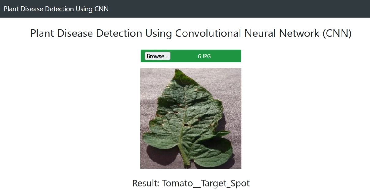
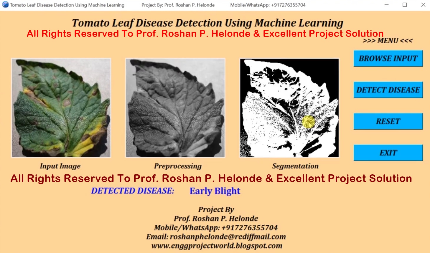
India is an agricultural country and soybean production is one of the major sources of earning. Due to the major factors like diseases, pest attacks, and sudden changes in the weather condition, the productivity of the soybean crop decreases. Automatic detection of tomato plant diseases is essential to detect the symptoms of tomato diseases as early as they appear on the growing stage. This project proposed a methodology for the analysis and detection of tomato plant leaf diseases using recent digital image processing techniques. In this project, experimental results demonstrate that the proposed method can successfully detect and classify the major tomato leaf diseases like Bacterial Spot, Early Blight, Leaf Curl Virus, Mosaic Virus, Healthy Leaf, etc.
We present a system where image analysis studies aiming at automated detection of disease that may be present in walnut by using image processing. In this work, we propose a convolution image processing model that has been trained with images of walnut. The project's aim is to build a fully automated walnut disease detection using convolutional neural network CNN in this categories into two category one is healthy walnut and secondly walnut affected with disease.
Determining of blood types is very important during emergency situation before administering a blood transfusion. Presently, these tests are performed manually by technicians, which can lead to human errors. Determination of the blood types in a short period of time and without human errors is very much essential. A method is developed based on processing of images acquired during the slide test. The image processing techniques such as Pre-processing, Segmentation, Thresholding, Morphological operations and Support Vector Machine are used. The images of the slide test are obtained from the pathological laboratory are processed and the occurrence of agglutination are evaluated. Thus the developed automated method determines the blood type using image processing techniques. The developed method is useful in emergency situation to determine the blood group without human error. The slide test consists of the mixture of one drop of blood and one drop of reagent, being the result interpreted according to the occurrence or not of agglutination. The combination of the occurrence and non occurrence of the agglutination determines the blood type of the patient.
Image processing is the recent growing technique in the world. It refers to the processing of digital images by means of a digital computer. Images play a major role in human perception. Image analysis is between image processing and computer vision. There are no clear boundaries for in continuum with image processing and computer vision. The useful paradigms for computerized process in determining the image is classified in to three types are low-level process: involve primitive operation such as image pre processing to reduce noise, image enhancement and image sharpening, mid-level: image segmentation and high-level: making sense of image recognized. Here image processing technique is used for medicinal purpose by extracting the features of herbal leaf and authenticating it medicinal qualities. Leaves play the major role for the classification of plants. The sample leaves are taken from various places, plants and shape. The image is captured and further work is carried out. Comparison of test sample image with reference not only requires an experienced but is subjective and prone to human errors. By applying advanced technique of image processing and utilizing the capabilities of the recent advanced computing and data/image storage facilities and the use of computer techniques for analyzing the shape, texture, color, aspect ratio, vein structure, entropy, compactness and so on. The aim of the work is to classify and authenticate the medicinal plant materials and herbs widely used for Indian herbal medicinal preparation. The quality and authenticity of these leaves are to be ensured for the preparation of herbal medicines. The medicinal plant leaves are thoroughly screened, analyzed and compared with the database to give the correct measures of the texture to which category the leaf belongs to. This method is adopted due to the mistaken of lookalike leaves using image processing technique the mistaken of look-alike leaves can be authenticated by various parameters of the leaves.
The lossless compression is that allows the original data to be perfectly reconstructed from the compressed data. Lossless compression programs do two things in sequence: the first step generates a statistical model for the input data, and the second step uses this model to map input data to bit sequences in such a way that probable. The main objective of image compression is to decrease the redundancy of the image data which helps in increasing the capacity of storage and efficient transmission. Image compression aids in decreasing the size in bytes of a digital image without degrading the quality of the image to an undesirable level. Image compression plays an important role in computer storage and transmission. The purpose of data compression is that we can reduce the size of data to save storage and reduce time for transmission. Image compression is a result of applying data compression to the digital image.
India is an agricultural country and soybean production is one of the major sources of earning. Due to the major factors like diseases, pest attacks, and sudden changes in the weather condition, the productivity of the soybean crop decreases. Automatic detection of soybean plant diseases is essential to detect the symptoms of soybean diseases as early as they appear on the growing stage. This project proposed a methodology for the analysis and detection of soybean plant leaf diseases using recent digital image processing techniques. In this project, experimental results demonstrate that the proposed method can successfully detect and classify the major soybean diseases like Soybean Rust, Powdery Mildew, Frogeye Leaf Spot, Downy Mildew, etc.
Malaria is an extremely infectious disease cause due to blood parasite of genus plasmodium. Malaria is a terrible disease in the hematological region causing millions of mortality; hence the fast diagnosing is the extreme requirement of era. Conventional microscopy, which is presently “the gold Standard” for malaria detection has occasionally proved ineffective as it takes lots of time and outcomes are complicated to reproduce. Since it poses a global health problem, automation of the evaluation method is of high significance. An image processing system is able to enhance outcomes of detection of malaria parasite cell. A variety of image processing techniques are used in the proposed method. The method proceeds in steps like image transformation, classification and feature extraction. This method assists to reduce time as well as afford the accuracy to detect malaria to certain extent. There are lots of methods to detect malaria, among them manual microscopy is considered to be "the gold standard". However because of the various steps essential in manual estimation, this diagnostic technique takes too much time. Malaria infections are detected manually by pathologists who observe the microscopic images of strained blood records on glass slides and calculate the contaminated blood cells. If sample size of patient is great, there is always a possibility to detect imprecisely. There is a chance to occur human error, so computer based classification using digital image processing methods gives better outcome than the manual diagnoses of Malaria. Intend of this work is to build up a detection method to correctly detected malaria parasites present in images. In the pre-processing stages digital image processing systems are used to obtain high-quality medical images. In this project, Image Processing is used to detect the existence of Malaria Parasite. In the proposed system, various steps are used such as image transformation, feature extraction and image classification.
Emotion Recognition is a recent research topic in the field of Human Computer Interaction Intelligence and mostly used to develop wide range of applications such as stress management for call centre employee, and learning & gaming software, In E-learning field, identifying students emotion timely and making appropriate treatment can enhance the quality of teaching. Main aim of HCI is to achieve a more natural interaction between machine and humans. HCI is an emerging field using which we can improve the interactions between users and computers by making computers more respond able to the user’s needs. Today’s HCI system has been developed to identify who is speaking or what he/she is speaking. If in the HCI system, the computers are given an ability to detect human emotions then they can know how he/she is speaking and can respond accurately and naturally like humans do. In this project methodology for emotion recognition from speech signal is presented. Here, some of acoustic features are extracted from speech signal to analyze the characteristics and behavior of speech. The system is used to recognize the basic emotions. It can serve as a basis for further designing an application for human like interaction with machines through natural language processing and improving the efficiency of emotion. In this format, energy, Mel Frequency Cepstral Coefficients (MFCC) has been used for feature extraction from the speech signal. Support Vector Machine (SVM) are used for recognition of emotional states. English datasets are used for analysis of emotions with SVM Kernel functions.
This project presents an iris coding method for effective recognition of an individual. The recognition is performed based on a mathematical and computational method. It consists of calculating the differences coefficients of overlapped angular patches from the normalized iris image for the purpose of feature extraction. Iris recognition belongs to the biometric identification. Biometric identification is a technology that is used for the identification an individual based on ones physiological or behavioral characteristics. Iris is the strongest physiological feature for the recognition process because it offers most accurate and reliable results. Iris recognition process mainly involves three stages namely, iris image preprocessing, feature extraction and template matching. In the pre-processing step, iris localization algorithm is used to locate the inner and outer boundaries of the iris. Detected iris region is then normalized to a fixed size rectangular block. In the feature extraction step, texture analysis method is used to extract significant features from the normalized iris image.
Reversible Data Hiding (RDH) techniques have gained popularity over the last two decades, where data is embedded in an image in such a way that the original image can be restored. Earlier works on RDH was based on the Image Histogram Modification that uses the peak point to embed data in the image. More recent works focus on the Difference Image Histogram Modification that exploits the fact that the neighboring pixels of an image are highly correlated and therefore the difference of image makes more space to embed large amount of data. In this project we propose a framework to increase the embedding capacity of reversible data hiding techniques that use a difference of image to embed data. The main idea is that, instead of taking the difference of the neighboring pixels, we rearrange the columns (or rows) of the image in a way that enhances the smooth regions of an image. Any difference based technique to embed data can then be used in the transformed image. The proposed method is applied on different types of images including textures, patterns and publicly available images.
Liver cancer is one of the most severe types ofcancerous diseases which is responsible for the death of many patients. Liver cancer images have more noises which is difficult to diagnose the level of the tumor. It is a challenging task to automatically identify the tumor from images because of several anatomical changes in different patients. The cancer is difficult to find because of the presence of objects with same intensity level. In this proposed system, fully automated machine learning is used to detect the liver tumor from input image. Region growing technique is used to segment the region of interest. The textural feature are extracted from Gray level co-occurrence matrix (GLCM) of the segmented image. Extracted textural features are given as input to the designed SVM classifier system. Performance analysis of SVM classification of liver cancer image is studied. This will be useful for physician in better automatic diagnosis of liver caner from input images.
Lung nodule prevalence is one of the highest of cancers, at 18 %. One of the first steps in lung nodule diagnosis is sampling of lung tissues or biopsy. These tissue samples are then microscopically analyzed. This procedure is taken once imaging tests indicate the presence of nodule cells in the chest. Lung nodule diagnosis using lung images. One of them is that doctor still relies on subjective visual observation. A medical specialist must do thorough observation and accurate analysis in detecting lung nodule in patients. Hence, there is need for a system that is capable for detecting lung nodule automatically from microscopic images of biopsy. This method will improve the accuracy and efficiency for lung nodule detection. The aim of this research is to design a lung nodule detection system based on analysis of microscopic image of biopsy using digital image processing. Microscopic images of biopsy are feature extracted and classified using support vector machine. This method is implemented to detection of lung nodule of lung samples.






.png)


